Students should refer to Cell Cycle and Cell Division ICSE Class 10 Biology notes provided below designed based on the latest syllabus and examination pattern issued by ICSE. These revision notes are really useful and will help you to learn all the important and difficult topics. These notes will also be very useful if you use them to revise just before your Biology Exams. Refer to more ICSE Class 10 Biology Notes for better preparation.
ICSE Class 10 Biology Cell Cycle and Cell Division Revision Notes
Students can refer to the quick revision notes prepared for Chapter Cell Cycle and Cell Division in Class 10 ICSE. These notes will be really helpful for the students giving the Biology exam in ICSE Class 10. Our teachers have prepared these concept notes based on the latest ICSE syllabus and ICSE books issued for the current academic year. Please refer to Chapter wise notes for ICSE Class 10 Biology provided on our website.
Cell Cycle and Cell Division ICSE Class 10 Biology
TOPIC-1
Cell Cycle and Cell Division
Quick Review
➢ All living beings are made up of one or more units called cells.
➢ The organisms made up of a single cell are called unicellular organisms. The organisms consists of many cells are known as multicellular organisms.
➢ The study of cell is called as Cytology. The term cell was given by Robert Hooke for the first time observed cork cells under a primitive microscope assembled by him.
➢ Cell Theory : The credit of formulation of cell theory is given to a German Botanist M.J. Schleiden and a German Zoologist T. Schwann who clearly outlined the basic features of cell theory that
(a) All living organisms are made up of cells.
(b) Cells always arise from the pre-existing living cells by division.
➢ Growth is a continuous process which occurs throughout the life of cell.
➢ Each and every cell in an organisms posses three essential part – Cell membrane, Cytoplasm and Nucleus.
➢ Cell division is necessary for proper growth, development and survival of organisms.
➢ Cell cycle is the sequence of events including growth and division, that cell undergoes from the time of its formation up to its division into daughter cells is called cell cycle.
➢ Cell cycle comprises of two phases – interphase and phase of division (mitosis and meiosis)
➢ Interphase is a series of changes that takes place in a newly formed cell and its nucleus before it becomes capable of division again. Therefore, it is also called intermitosis.
➢ Interphase of dividing cell has three stages – G1 , S and G2.
➢ Interphase also know as biosynthetic phase in which cell duplicates its cell organelles & replicates its DNA.
➢ G1 phase is characterized by the synthesis of RNA and non-histone proteins. Cell growth occur and substances are produced which inhibit or stimulate the onset of next S – phase.
➢ S – phase follows the G1 – phase. It is characterized by the replication of DNA and the chromosome are completely duplicated.
➢ G2 – phase is the period in which centrioles, mitochondria, Golgi bodies and other cytoplasmic organelles are doubled.
➢ M or D phase is the phase when the cell enters the prophase stage of cell division.
➢ G1 , S , G2 and M – phase collectively form the cell cycle.
➢ Mitotic ( M – phase ) consists of karyokinesis (division of nucleus) and cytokinesis (division of cytoplasm).
➢ Mitosis was first observed by Strasburger in plant cells & by flaming in animal cells.
➢ Karyokinesis comprises of four phases – prophase, metaphase, anaphase and telophase.
➢ Prophase is the longest phase of division. Replicated chromosomes each with sister chromatids condense and become visible. Nuclear membrane along with nucleolus disappear.
➢ In metaphase chromosomes arranged themselves in the equator of the spindle to form the equatorial plate. Each chromosome is attached to the spindle fibres by its centromere.
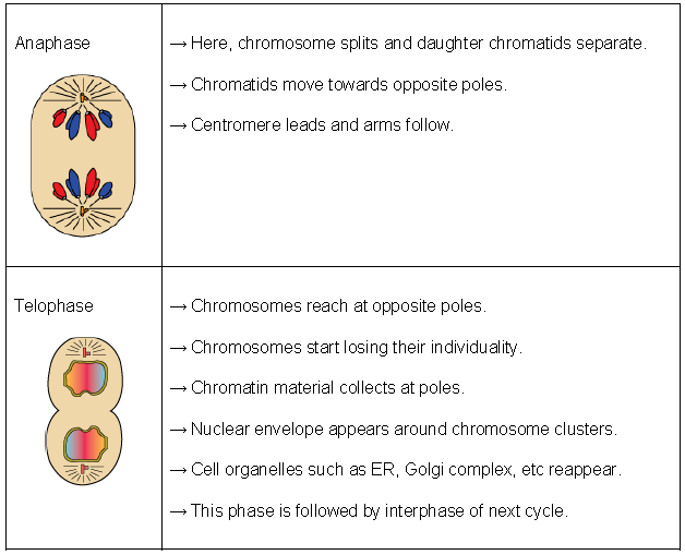
➢In anaph ase the centromere divide there by separating into two sister chromatids or newly formed chromosomes. Sister chromatids of each chromosome move towards the opposite poles of the spindle.
➢ Telophase is the phase in which nuclear membrane is reformed around each group of chromatids, now called chromosomes at each pole. Two nuclei are thus formed.
➢ Karyokinesis is followed by cytokinesis.
➢ Mitosis results in the formation of identical cells. Daughter cells have same genetic constitution quantitatively and qualitatively as the original cell.
➢ It brings about the reproduction in unicellular organisms.
➢ It is necessary for growth, maintenance and repair in multicellular organisms.
Mitosis in Animal cell Mitosis in plant cell
1. Aster are formed. 1. Asters are not formed.
2. Cytokinesis occurs by furrowing of Cytoplasm. 2. Cytokinesis occurs by cell plate formation.
3. Occurs in most tissues throughout the body 3. Occurs mainly at the growing tips & sides.
➢ Meiosis is the cell division which occurs in sex cells and results in the formation of four daughter cells. The daughter cells are quantitatively and qualitatively different from mother cells.
➢ The whole process consists of two successive coordinated divisions called meiosis – I and meiosis – II.
➢ During meiosis – I the number of chromosomes reduced to half while the meiosis – II division is the simple mitotic type.
➢ Meiosis maintains the same chromosomes number through successive generation of species.
➢ The most significant of Meiosis is that the number of chromosomes in the sex cells is halved.
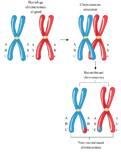
➢ It helps to produce the new recombination of characters as a result of crossing – over ( exchange of genetic material between two homologous chromosome.
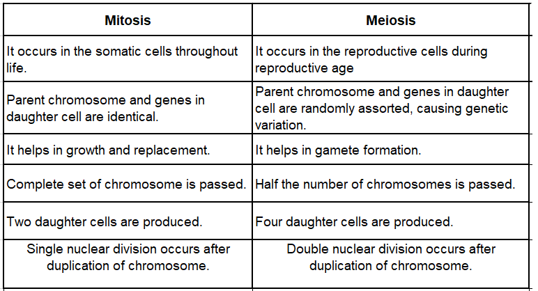
➢ Major differences in Mitosis and Meiosis are
➢ Mitosis Meiosis
(a) Mitosis takes place in the somatic cells. It occurs either in the reproductive cells or at the time of germination of zygote.
(b) It is a single division which produces two cells. It is a double division. It gives rise to four cells.
(c) The number of chromosomes remains the same after mitosis. The number of chromosomes reduced to half After meiosis.
(d) No crossing over takes place. Crossing over takes place in prophase – I
TOPIC-2
Structure of Chromosome
Quick Review
➢ Chromosomes are rod-shaped or thread like deeply stainable condensed chromatin fibres which are hereditary vehicles.
➢ Hofmeister(1848) first observed the chromosome. The name chromosome was proposed by W. Waldeyer (1888).
➢ Role of chromosome in hereditary process was discovered by Morgan (1933).
➢ A set of paired chromosomes or two sets of chromosomes is known as diploid. A set of unpaired chromosomes or a single set of chromosomes is said to be a haploid.
➢ Each species has a definite number of chromosome.
➢ Each chromosome consists of 2 units or arms, called chromatids attached at a point called primary constriction or centromere.
➢ Depending on the location of centromere, chromosomes are classified into four types –
(i) Telocentric – Centromere at one tip.
(ii) Acrocentric – Centromere is just below the tip.
(iii) Sub-metacentric – Centromere is in between the center and tip of the chromosome.
(iv) Metacentric – Centromere is in the middle.
➢ A chromatin fibre is a continues linear DNA double strand associated with proteins of two types – Basic histones and acid or neutral non-histones. It also contains some RNA and some enzymes such as DNA and RNA polymerases.
➢ The term chromatin means “coloured material “. The chromatin occurs in a non-dividing nucleus as fine filaments termed as the chromatin fibres.
➢ A chromosome consists of two identical halves, the chromatids, held together at one point, the centromere.
➢ Double helical model to explain the structure of DNA was given by Watson & Crick in 1953.
➢ The organisms which contain a segment of foreign DNA or gene are known as transgenic organisms.
Know the Terms
➢ Amitosis : Cell divides without spindle formation.
➢ Disjunction : It is the separation of homologous chromosomes during cell division.
➢ Non-disjunction : It is the non-separation of homologous chromosomes during anaphase – I of meiosis – I.
➢ Congression : Chromosomes fibres contract and bring the chromosome over the equator.
➢ Mitogens : The agents which stimulate the cell division are called mitogens. E.g. Cytokinins and some steroids.
➢ Mitotic poisons : There are some chemicals which inhibit cell division e.g. azides, cyanides, colchicine.
➢ Intranuclear mitosis and pre-mitosis : In protists, fungi and algae the nuclear envelop does not degenerate during mitosis. Instead spindle is formed inside the nucleus.
➢ Centromeric Index : It is the ratio of lengths of the two arms of chromosome.
➢ Allosomes : These are sex chromosomes whose presence, absence and particular form determines the sex of the individual.
➢ Autosomes : Chromosomes other than sex chromosomes are called autosomes.
➢ Homogametic : Individuals having homomorphic sex chromosomes produce similar gametes. E.g. human female
➢ Heterogametic : Individuals with heteromorphic sex chromosome produce two types of gametes. E.g. Human male.
➢ Karyotype : Karyotype is a chromosome complement of a cell / organism providing description of various aspects of all the chromosomes like number, relative size, position of centromere, length of arms and satellite.
➢ Satellite / Trabant : A satellite chromosome or SAT chromosome has a chromosome segment that is separated from the main body of the chromosome by such a secondary constriction.
Cells: An Overview
Diverse forms of living organisms are present in our surroundings. Like ourselves, all of them are made up of tiny structures called cells. Cells are the building blocks of life. They are the basic structural and functional units from which life takes shape. A cell is the smallest living entity in a living organism.
How cells are formed
In 1838 & 1839, the two German scientists Matthias Schleiden (1838) and Theodor Schwann (1839) proposed the cell theory and formulated that all plant and animal tissues are made up of cells. They, however, were unsuccessful in explaining how new cells are formed. Later, in 1855, Rudolf Virchow further expanded the cell theory by suggesting that all cells arise from pre-existing cells. The cell theory states that:
• All living organisms are composed of cells and products of cells.
• Cells are the basic units of structure and function in an organism.
• All cells arise from pre-existing cells.
Know Your Scientist

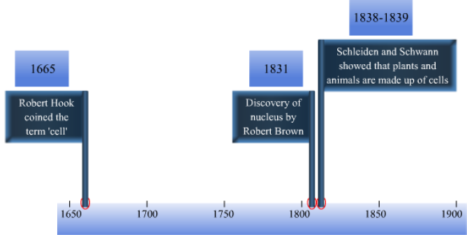
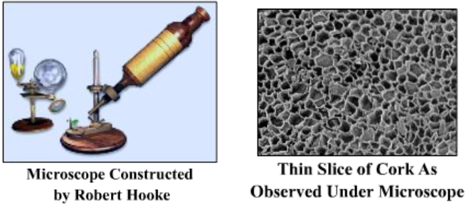
The term ‘cell’ was introduced by Robert Hooke in 1665 after observing the cellular structure of cork (a substance obtained from the bark of a tree). While examining a thin slice of cork under a compound microscope, Hooke observed many small compartments resembling honeycombs. These he termed as cells.

In 1831, Robert Brown discovered the presence of nucleus in the centre of a plant cell.
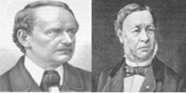
Theodor Schwann (1810-1882) and Matthias Schleiden (1804-1881)
In 1838, Matthias Schleiden, a German physiologist, discovered that all plant tissues are made up of cells, i.e., cells are the fundamental units of all plants. In the next year(1839), Theodor Schwann, a German physiologist, discovered that all animal tissues are made up of cells, i.e., cells are the fundamental units of all animals.
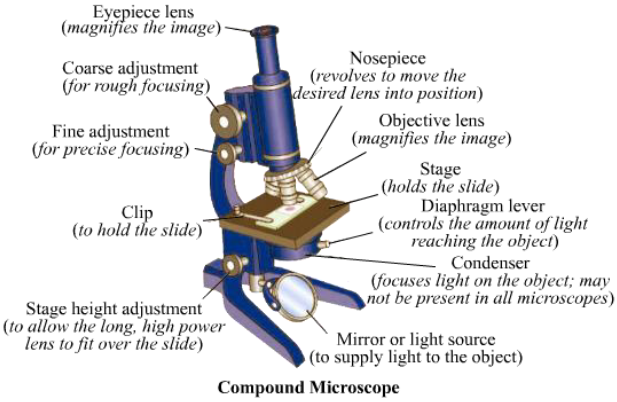
Invention of Microscope
Cells are very small living entities that are not visible to the naked eyes. The invention of microscopes hence played the key role in the discovery of cells.
Simple Microscopes
• First simple microscopes were constructed by Antony van Leeuwenhoek (1632-1723).
• They consisted of single biconvex lens.
• Their magnifying power was up to 200 times.
Compound Microscopes
• These were first constructed by Robert Hooke (1635-1703).
• He developed the compound microscope using two lenses for increasing the magnifying power.
• He examined a thin slice of cork under it and observed tiny, box-like compartments, that he named ‘cells’.
The modern ordinary compound microscope has greatly improved in design and magnification power (up to 2,000 times).
Electron Microscopes
• The invention of electron microscope has led to great advancements in the study of cells.
• Electron microscopes use beams of electrons which are bent by magnets to magnify the images.
• They can magnify an object up to 200,000 times.
Cell-The Basic Unit of Life
Properties of Living Cells
Some important properties of living cells are as follows:
• Generally, a cell is so small that it is not visible to the naked eye.
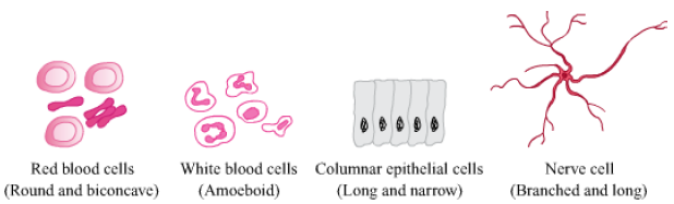
• Cell shape and size vary both within an organism and between different organisms. The shape and size of a cell is related to the specific function it performs.
• All living cells exhibit certain basic properties like respiration, growth and metabolism.
• Nerve cells are some of the longest cells.
Examples of cells with different shapes and sizes
Did You Know?
• The smallest unicellular organism we know is the Mycoplasma, a type of bacteria. Its diameter is 0.1 μm.
• There are more red blood cells in our body than any other type of cell.
Solved Examples
Medium
Example 1: Illustrate how the shape and size of a cell is related to the specific function it performs.
Solution: Different types of cells with different shapes and sizes are present in our body. A cell’s shape and size are relevant to the specific function it performs. The irregularly shaped white blood cell is a case in point. A white blood cell protects the body by killing harmful foreign bodies. Whenever it encounters any antigen, it changes its shape accordingly and engulfs the antigen. Thus, the shape of the white blood cell is directly related to the function it performs.
Classification of Cells
Based on the number of cells: Unicellular and multicellular

As you now know, a cell is the smallest living entity capable of independent existence. There are certain organisms that are made up of only a single cell; such organisms are known as unicellular organisms. Examples of unicellular organisms include Amoeba and yeast. All other organisms (i.e. those made up of more than one cell) are known as multicellular organisms. Examples of multicellular organisms include humans, plants and animals.
• Multicellular organisms can perform a variety of tasks efficiently due to division of labour. This gives the organisms a wide range of adaptabilities to survive.
• In multicellular organisms, dead cells play an important role. For example, the dead epidermal cells in animal skin protect the underlying cells.
Division of labour
Divisio12701n of labour refers to the specialized roles of the different organs present in a multicellular organism. All organs, tissues or cells of a multicellular organism cannot carry out all the functions.
Each of them is evolved to carry out a specific set of tasks. Each organ system coordinates with the others to perform the activities required for life. This division of labour minimizes the load of carrying out all the functions and, consequently, it allows the organs to operate efficiently.
Concept Builder
Let us understand this concept of division of labour using the example of a cricket team. As you know, in a cricket team, some members specialize in batting while some specialize in bowling. Each member is assigned a specific set of functions in the team. More often than not, the team that wins a game is one whose members perform their specific tasks efficiently.
In the same way, the different organ systems in the human body are assigned different functions. For example, the digestive system is assigned to carry out digestion, while the excretory system is assigned to carry out excretion.
This is division of labour. Ultimately, a healthy body is one in which the different organ systems perform their respective functions properly.
Classification of Cells
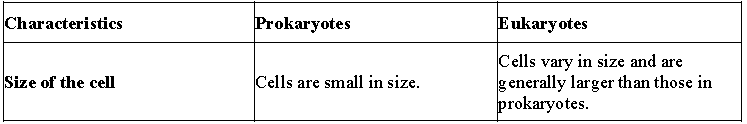
Based on the cellular complexity: prokaryotes and eukaryotes
This type of classification is based on the sub-cellular organization of a cell.
The given table lists the characteristic features of prokaryotes and eukaryotes.

Solved Examples
Medium
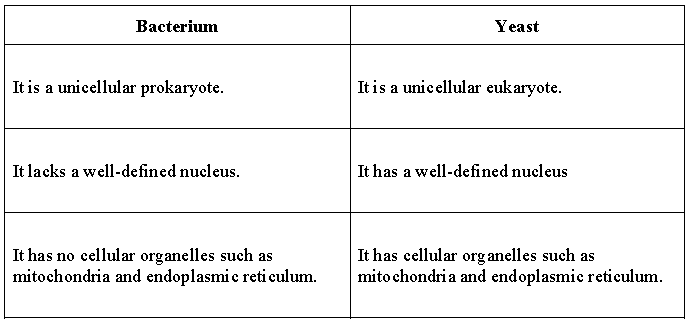
Example 2:
Distinguish between bacteria and yeast.
Solution:
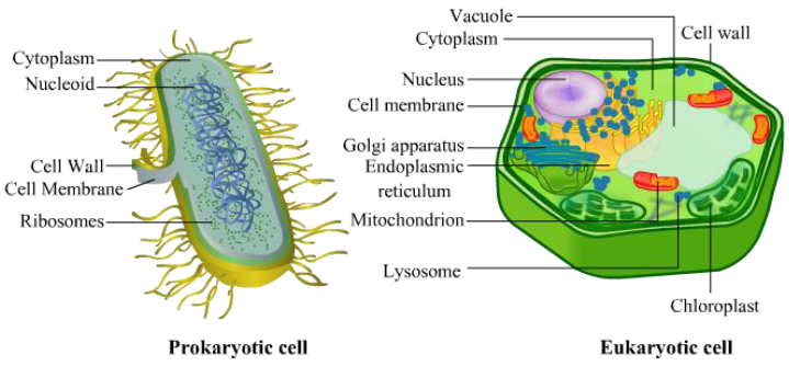
Structure of Eukaryotic and Prokaryotic Cell
Difference between Plant Cell and Animal Cell
Cell Division
Cells undergo division to form new cells. These new cells are used to grow, replace old, dead and injured cells, and to form gametes required for reproduction. There are two types of division a cell undergoes –
Mitosis – Each cell divides to form two daughter cells. The daughter cells have the same chromosome number as the mother cell. Meiosis – This type of division is shown by specific cells of the reproductive organs or tissues in animals and plants. These cells divide to form gametes, which after fertilisation give rise to new off springs. In meiosis, four cells are produced from a single cell and the new cells have half the chromosome number than the mother cell.
Cell Organelles: Their Structure and Functions
We know that cell is the basic structural and functional unit of life. But what is present inside a cell? How does it perform its various functions?
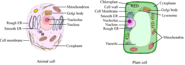
A cell consists of three essential parts: cell membrane, cytoplasm and nucleus. Let us know more about these parts.
Cell membrane:
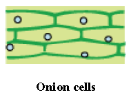
Take a peel of onion by separating it from the fleshy portion. Add a drop of methylene blue on a slide containing the peel, put cover slips, and observe it under a microscope. What do you observe? Note your observations and draw a diagram of the structure you have observed.
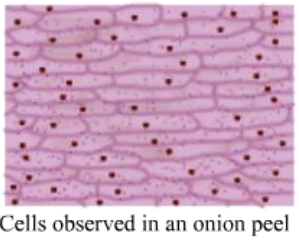
You will observe brick-like cells placed one over the other with a round ball-like nucleus at the centre. The boundary of the onion cells is known as the cell membrane or plasma membrane and it is covered by another thick layer called the cell wall.
The cell membrane is porous in nature and helps in the inward and outward movement of substances. However, it is selectively permeable in nature, which means that it allows the in and out movement of only certain substances.
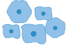
Let us now observe how animal cells look under a microscope. Given below is an image of human cheek cells as observed under a microscope.

Do you notice any difference between these cells and onion cells? The cell membrane in this case is not surrounded by any other layer!
Hence, in plants, cell membrane is surrounded by another layer known as cell wall whereas animal cell contain only cell membrane.
Cell wall:
The cell wall is an additional protective, rigid structure present outside the cell membrane. It is present only in plant cells. It protects them from heat, humidity, pressure, etc. It also gives the plant cells their characteristic shape and rigidity. It is freely permeable in nature.
Cytoplasm: The jelly-like substance present between the cell membrane and the nucleus is called the cytoplasm. It is an important component of the cell as various cell organelles such as mitochondria, ribosomes, etc. are present in it.
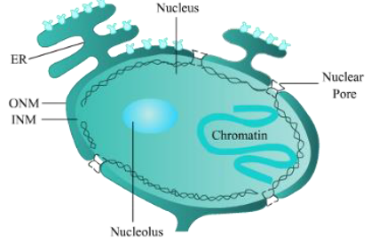
Nucleus:
Nucleus is a spherical structure, which is generally present at the centre of the cell.
Parts of Nucleus
• Nuclear membrane: The nucleus is enclosed by a double-walled cellular membrane called the nuclear envelope. The nuclear envelope separates the contents of the nucleus from the cytoplasm. The nuclear membrane is pierced with holes known as the nuclear pores. These pores allow the nucleus to communicate with the rest of the cell.
• Nucleolus: It is a spherical structure found inside the nucleus. It plays an important role in protein synthesis.
• Nucleoplasm: The nucleus contains a semi-fluid substance known as nucleoplasm or karyoplasm. It holds the nucleolus and the suspended chromatin.
• Chromatin network: The nucleus contains the genetic material of an organism in the form of a network of chromatin. This chromatin gets folded and coiled to form chromosomes. Let us study the components of nucleus by this video.
Cell membrane, cytoplasm, and nucleus form the basic components of the cell.
Some interesting facts:
• Do you know that the red blood cells of the human body do not have a nucleus?
• Paramecium is a unicellular organism having two nuclei.
• Some muscle cells in humans have a large number of nuclei.
CELL ORGANELLES
These are the living parts of a cell that have definite shapes, structures and functions. Let us explore all the cell organelles found in a cell.
Vacuole: When you observe an onion peel under the microscope, you will observe large empty structures in the cells. Do you know what these structures are? These empty structures are called vacuoles. These vacuoles are larger in plant cells than in animal cells.
Vacuoles are membrane-bound structures, which are believed to store substances in cells. In plant cells, vacuoles are large in size, while in animal cells vacuoles are small. The table given below lists some functions of vacuoles. The membrane of vacuoles is called tonoplast.
Functions of vacuoles:
• They help in the removal of unwanted structural debris.
• They store all the waste products of cells.
• In Amoeba, food vacuoles store food.
Plastids:
Take a peel of the Tradescantia leaf and observe it under the microscope. You will find coloured bodies in the cytoplasm of the leaf cells. Do you know what these are? These are called plastids. The green coloured plastids in the cell are known as chloroplasts. They are responsible for the green colour of the leaves. They carry out the process of photosynthesis and help plants prepare their own food.
Do you know that some plastids are specialized to store starch, proteins, and lipids?
Plastids are major organelles found in plant cells and algae. There are two major types of plastids, namely Chromoplasts and leucoplasts.
Chromoplasts are coloured plastids, while leucoplasts are white or colourless plastids. Chromoplasts contain coloured pigments like carotene (orange), xanthophylls (yellow) etc. These pigments are responsible for the colour of plants. Unlike chromoplasts, leucoplasts lack pigments.
Chloroplasts are plastids containing the pigment called chlorophyll. A chloroplast is enclosed by two lipid membranes. They are called the kitchen of the cell.

The inner matrix is called the stroma. Thylakoids are the sub-organelles arranged in stacks within the stroma to form grana. Plastids also contain their own DNA and ribosomes.
Functions of plastids
• They carry out the process of photosynthesis.
• They contribute to the colour of leaves, flowers etc.
Endoplasmic Reticulum
Endoplasmic reticulum, or ER, is an interconnected network of membranous structures like tubules, vesicles, and cisternae. Cisternae are the flattened disc-like membranous structures. Tubules are tubular in shape, while vesicles are sac-like structures.
There are two types of endoplasmic reticulum, namely smooth endoplasmic reticulum (SER) and rough endoplasmic reticulum (RER). When ribosomes get attached to the surface of smooth endoplasmic reticulum, it becomes rough endoplasmic reticulum.
The basic functions of endoplasmic reticulum are
• To help in protein and lipid synthesis.
• To provide internal support to the cells.
• To provide transportation pathway within the cells.
Ribosomes
Ribosomes are the small granular structures that help in the protein synthesis. Hence, they are also known as the “protein factories” of the cell.
Golgi Apparatus
Golgi apparatus have the membrane-bound, sac-like structures called cisternae and some small vesicles. They are arranged parallel to each other in stacks. They were discovered by Camillo Golgi in 1898. Golgi body is usually composed of five to eight cisternae in stacks. Some functions of the Golgi apparatus are enlisted below.
Functions of Golgi apparatus
• It involves the transport of lipids in cells.
• It involves the formation of lysosomes.
• It is involved in the synthesis of cell wall in the plant cell.
• It is involved in the modification, sorting and packaging of proteins.
The golgi apparatus present in the plant cell are called dictyosomes. They are small, unconnected and more in number as compared to the animal cell.
Mitochondria
Mitochondrion is a membrane-enclosed organelle found in eukaryotic cells.
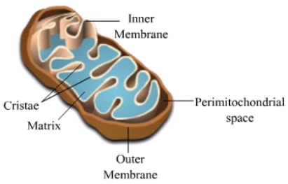
Mitochondria are responsible for the production of most of the energy (or ATP) in cells. Therefore, mitochondria are also known as the power house of cells. A mitochondrion is composed of two lipid membranes, enclosing the matrix. The inner membrane gets folded to form numerous cristae. Cristae are the main site for ATP production. Mitochondrial matrix contains mitochondrial DNA and ribosomes.
Functions of mitochondria
• They produce energy required for cells in the form of ATP.
• They also regulate the free calcium ion concentration in the cytosol.
• They participate in apoptosis or programmed cell death.
Lysosomes
Lysosomes are the membrane-bound vesicles, which contain digestive (hydrolytic) enzymes. They digest a variety of substances including worn out organelles, food particles, viruses, and bacteria. They are also known as ‘suicide-bags’ of cells as they burst out and release hydrolytic enzymes in the cytosol, causing destruction of the damaged or injured cells.
Functions of lysosomes
• They digest macromolecules by phagocytosis. So, they provide protection to the cell against foreign substances.
• They also take part in auto-cell lysis.
Centrosome Centrosome is found exclusively in animal cells. It lies very close to the nucleus. It contains two cylindrical structures called centrioles.
Both centrioles in a centrosome lie perpendicular to each other. Centrioles have a cartwheel-like organisation.
The centriole has a role in cell division.
Cell Inclusions
Cell inclusions are the result of various chemical reactions that take place inside the cell, either in the cytoplasm or in the vacuole.
Cell inclusions may be the food products like starch or oil globules or the waste materials like gums, resins, tannins, and latex.
Observation of Plant and Animal Cell
Let us perform an experiment to understand how does a cell look like under microscope?
Take an onion and cut it into two halves. Peel off a transparent piece of skin from the inner layer of onion with the help of a forceps. Place this transparent skin on a slide and add a drop of iodine solution to it. Carefully place the cover slip on the slide. Wipe off the excess amount of iodine solution from the slide with the help of tissue paper. Observe the slide under microscope. Carefully have a look on the shape of onion cell.

Observation:
You can see several rectangular cells, each with a small, spherical nucleus in it. These are called epidermal cells that are found on the surface of the plant body.
Cells are basic unit of life that is capable of doing all the required biochemical processes that a normal cell requires to do in order to live. The basic need for the survival of all living organism are same. All living organisms need to digest food (to obtain energy), respire and to get rid of metabolic wastes. Who does all these functions of the body?
It is the cell that carries all these metabolic function in body. Hence, cell are called functional unit of life.
Observing Animal Cell
Take a cotton bud and gently rub it along inside of your cheek. Smear the cotton bud onto a slide. Add a drop of methylene blue on to the smear. Carefully place a covers slip on top of the slide. Now observe the slide under microscope.
(Note: Methylene blue is a dye used to stain and view animal cells)
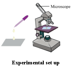
Observation
You can see several polygonal cells lying here and there on slide. This represents the structure of animal cell.
Differences between Plant and Animal Cell

Nucleus and Chromosomes
Nucleus

• Every cell has a nucleus, except some such as the RBCs of mammals and the sieve tube cells in vascular plants. A cell usually has one nucleus, except some variations.
• Nucleus is bound by a nuclear envelope which consists of two membranes with perinuclear space (10 − 50 nm) between them
• Perinuclear space acts as a barrier for the flow of materials between the inside of the nucleus and the cytoplasm. So, to facilitate the transfer of RNA and proteins, nuclear pores are present. Nuclear pores are formed at places where the two membranes fuse.
• Nuclear matrix (Nucleoplasm consists of the nucleolus and the chromatin.)
• Nucleolus (pl. nucleoli): Site of active rRNA synthesis; not bound by membrane; its contents are continuous with the nucleoplasm
• Chromatin: Loose, indistinct network of nucleoprotein fibres (present in the interphase)
• Chromosomes: Chromatin structures develop into distinct chromosomes during cell division.
• Contains DNA, histone proteins, non-histone proteins, and also RNA
• DNA is distributed among 23 pairs (46) of chromosomes
• A chromosome has a primary constriction called centromere. On the sides of the centromere, disc-shaped kinetochores are present.
Classification of Chromosomes
• Based upon the position of the centromere, chromosomes are of four types:
• Metacentric − centromere located in the middle, forming two equal arms of the chromosome
• Sub-metacentric − centromere located slightly away from the middle, resulting in one arm being longer than the other
• Acrocentric − centromere located close to the end, resulting in one arm being extremely longer than the other
• Telocentric − centromere located at the terminal point
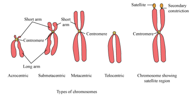
• Satellites: Small fragments that appear due to the non-staining secondary constrictions present at a constant location on the chromosomes
DNA and Its Structure
Deoxyribonucleic acid or DNA is a macromolecule found inside the nucleus. It has a double helical structure, similar to a ladder, which is twisted at both ends. A DNA molecule is made up of repeating units of nucleotides. Each nucleotide is made up of three components :

• A pentose sugar (ribose)
• Phosphate group
• A nitrogen base
There are four nitrogenous bases found in DNA. These are :
• Adenine (A)
• Guanine (G)
• Cytosine (C)
• Thymine (T)
Adenine pairs with Thymine with the help of two hydrogen bonds, while Guanine pairs with Cytosine with the help of three hydrogen bonds.
Each DNA molecule has a property to duplicate, or replicate, itself. This replication process takes place during mitosis, in which the helical structure of DNA gets open at one end and the free strands give rise to new, complementary strands.
Genes
A gene is a functional unit of DNA. It is located on a chromosome and controls the development of one or more traits through proteins encoded by it. It is the basic unit through which the genetic information is transferred from parent to their offspring. Every person has two copies of each gene, one inherited from each parent. Genes can acquire mutations in their sequence that lead to different variants, known as alleles, in population.
Phases of Cell Cycle
Cell Cycle
• The sequence of events by which a cell duplicates its genome, synthesises other cell constituents, and eventually divides into two daughter cells.
• The events of the cell cycle are under genetic control.
Phases of Cell Cycle
• Duration of the cell cycle varies from organism to organism, and from cell to cell. Duration of the cell cycle in humans is 24 hrs, and in yeast is 90 min.
• Phases of the cell cycle are:
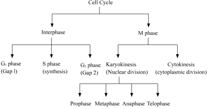
• Interphase represents the phase during which a cell prepares itself for division, grows and DNA is replicated in it. Interphase occupies more than 95% of the duration of the cell cycle.
• It is divided into three phases − G1, S and G2.
• G1 (Gap 1) phase is the phase between the end of mitosis and the initiation of DNA replication. The growth of the cell takes place in this phase.
• S (synthesis) phase is the phase of DNA replication. DNA content doubles (from 2C to 4C), but chromosome number remains the same. (remains diploid; 2n only). In this phase, the centriole also duplicates in animal cells.
• G2 (Gap 2) phase is the phase in which the proteins needed for mitosis are synthesised. Cell growth continues in this phase.

• G0 phase is the quiescent stage in which the cells that do not divide further enter after exiting from the G1 phase. In this phase, the cells do remain metabolically active, but do not proliferate unless required.
• M phase represents the phase where the cell actually divides. It starts with nuclear division and ends with the division of the cytoplasm.
Significance of Cell Division
• It is the mean of asexual reproduction in unicellular organisms.
• It is essential for the growth of a single celled zygote into a whole new multicellular organism.
• It helps in the repair of injuries and worn out tissues.
• It replaces dead cells of the body and thus is essential for growth of organism.
• In sexual reproduction, meiosis occurs. This type of cell division not only results in production of gametes, but also brings new combinations of genes, thus resulting in variations among a population. This also leads to evolution of a species.
Mitosis – Events and Significance
Mitosis (M Phase)
• Also called equational division as number of chromosomes in parent and progeny remain the same
• Mitosis (M phase) is divided into 4 stages.
• Prophase (1st stage)
• Metaphase
• Anaphase
• Telophase (Last stage)
Cytokinesis
• Cyto (cell) kinesis (division) follows the process of karyokinesis (nuclear division).
• Process of cytokinesis in plant cell:
• New cell wall begins to form with precursor cell plate equivalent to middle lamella.
• This new cell wall grows outwards.
• Organelles such as mitochondria and plastids also get distributed between two daughter cells at the same time.
• Process of cytokinesis in animal cell:
• Furrow appears in the cell membrane.
• This furrow deepens and joins in the centre to divide the cytoplasm into two.
• If cytokinesis does not follow karyokinesis, then cell becomes multinucleate, leading to the formation of syncytium
Significance of mitosis
• Results in formation of diploid genetically identical daughter cells
• Growth in multicellular organisms takes place by mitosis.
• Cell repair and replacement of worn out tissues
• Maintenance of nucleo-cytoplasmic ratio
• Vegetative reproduction in plants takes place by mitosis.
Meiosis and Its Significance
Meiosis
• It is a type of cell division that produces gametes.
• It occurs in the reproductive organs of various organisms. In humans, it occurs in testis and ovary to produce sperm and ova, respectively, whereas in plants, it occurs in anthers and ovules to produce pollen grains and egg, respectively
. Important characteristic:
• In normal body cells, or somatic cells, the chromosomes are present in the form of pairs; each set of a pair comes from each parent. Such a cell is said to be diploid, represented as 2n.
• During meiosis, the number of chromosomes is halved, so that the daughter cells thus formed contain single set of the chromosome pair. The cells thus formed (or gametes) are said to be haploid, represented as n.
• This reductional nature of meiosis is important to maintain the genetic makeup of an organism undergoing sexual reproduction. In sexual reproduction, the haploid male and female gametes produced through meiosis get fused together during fertilisation. As a result, the chromosome number in the zygote remains equal to that of the parents, and thus the diploid nature is restored.
Significance of meiosis:-
• Chromosome number is halved.
• It helps in mixing up of genes. This occurs in two ways:
• The maternal and paternal chromosomes mix at the time of first division, when they separate from homologous pairs. • Cross joining : When maternal and paternal chromosomes separate, chromatid material often gets exchanged between the members of homologous pair, resulting in genetic recombination.
Differences between Mitosis and Meiosis