Students can refer to the following Sample Paper ICSE Class 10 Biology Set B with Answers provided below based on the latest syllabus and examination guidelines issued for ICSE Biology. All specimen papers have been prepared covering all chapters given in ICSE Biology book for Class 10. You should also refer to ICSE Class 10 Biology Solutions.
Sample Paper ICSE Class 10 Biology Set B
Class: Time: 2Hrs.
Max. Marks:80
BIOLOGY
Instructions
You will not be allowed to write during the first 15 minutes.
This time is to be spent in reading the Question Paper.
The time given at the head of this Paper is the time allowed for writing the answers.
Attempt all questions from Section I and any four questions from Section II.
The intended marks for questions or parts of questions are given in brackets [ ].
SECTION I (40 Marks)
Attempt all questions from this Section
Q.1.
a. Name the following: [6×1=6]
i. The fluid entering PCT after ultrafiltration.
ii. The chest pain caused due to insufficient supply of blood to heart muscles.
iii. The pigment that causes yellow coloration of urine.
iv. The largest WBC present in human blood.
v. The cords that keep tricuspid valve in position.
vi. The process by which cells adsorb water.
b. Fill in the blanks: [6×1-=6]
i. The muscles arising from the projection of the ventricle are called ————.
ii. Haemoglobin is present in the ———— region of RBC.
iii. —————- is the smallest WBC.
iv. Chromosomes are duplicated during ——- phase of cell cycle.
v. ———— is the site of dark reaction for photosynthesis in chloroplast.
vi. The average life span of RBC is about ————–.
c. State whether the following statements are True or False. If false, correct it by changing only one word. [6×1=6]
i. Guttation occurs through stomata.
ii. Eosinophil responds to basic dye.
iii. The protein part of Haemoglobin is haemin.
iv. Pulmonary artery carries pure blood.
v. Crossing over can occur only between sister chromatids.
vi. Abnormal increase in the number of RBC is called erythropenia.
d. Differentiate between the following pairs: [3×1=3]
i. Neutrophil and Basophil [function]
ii. Leukemia and Leucopenia [condition]
iii. Nucleoside and Nucleotide [composition]
e. Arrange the following terms in a logical sequence: [4×1=4]
i. Venacava, right auricle, lungs, aorta, left auricle, right ventricle, left ventricle.
ii. Liver, hepatic vein, stomach, hepatic portal vein, posterior vena cava.
iii. Afferent arteriole, renal vein, secondary capillary network, glomerulus, efferent arteriole.
iv. Bowman’s capsule, proximal convoluted tubule, distal convoluted tubule, loop of Henle, collecting duct.

g. Define the following terms: [4×1=4]
i. Diapedesis
ii. Osmotic pressure
iii. Ultrafiltration
iv. Micturition.
h. Observe the schematic diagram of human blood circulatory system and answer the following questions:

i. Which part represents the heart? Mention the number. Give reason to support the answer.
ii. Which numbers represent the following respectively?
a. Aorta
b. Hepatic portal vein
c. Pulmonary vein
d. Renal vein
e. Stomach
iii. What is double circulation?
i. Differentiate between artery and vein with the help of labelled diagrams.
PART II
ANSWER ANY FOUR QUESTIONS
Q.2.
a. Draw a neat labelled diagram of the L.S. of kidney. [3]
b. Schematically represent the mechanism of clotting of blood. [3]
c. Give reason for the following statements: [4]
i. Reabsorption of glomerular filtrate is called selective absorption.
ii. Lymph does not contain RBC and platelets.
iii. Ventricles have thick muscular walls.
iv. Excessive transpiration causes wilting.
Q.3.
a. Observe the diagram and answer the following questions

i. Label the parts 1 to 8
ii. Mention the function of any four parts.
iii. Why is 1 larger than 2?
iv. How is blood supply to the kidney tubules arranged?
b. The diagram given below represents the human heart in one phase of its activity. Study the same and answer the following questions:

i. Name the phase.
ii. Which part of the heart is contracting in this phase? Give a reason to support the answer.
ii. Name the parts numbered 1 to 6.
iii. Which type of blood flows through the parts marked 1 and 2 respectively?
iv. How many valves are closed in this phase?
Q.4.
a. List any four physical properties of human urine. [2]
b. Explain ABO system of blood grouping.
c. Given below is the figure of certain organs and associated parts in the human body. Observe the diagram and answer the following questions:

i. Name the organ system shown in the diagram.
ii. Name the parts numbered 1 to 5.
iii. Name the structural and functional unit of the part marked 1.
iv. Name the two main organic constituents of the fluid that flows down the part labelled 3.
v. Name the two major steps involved in the formation of the fluid that passes down the part labelled 3.
Q.5.
a. Explain hepatic portal system. [3]
b. Differentiate between pulmonary circulation and systemic circulation.
c. Study the diagram given below and answer the following questions:

i. Name the region in the kidney where the above structure is present.
ii. Name the parts labelled 1 to 4.
iii. Name the stages involved in the formation of urine.
iv. What is the term given to the process occurring in 2 and 3? Explain the processes.
Q.6.
a. Name the blood groups referred to as universal donor and universal recipient. Why are they referred to so? [2]
b. Explain briefly the role of the following structures in this process. [5]
i. guard cells
ii. Cuticle
iii. Chlorophyll
iv. Stomata
v. Xylem
c. Observe the given experiment and answer the following questions:
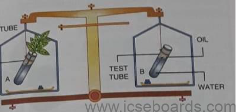
i. Name the process intended for study.
ii. When the weight of the test tubes is taken before and after the experiment, what changes are observed? Justify.
iii. What is the purpose of putting oil in the test tube?
Q.7.
a. Shown below are four stages of a certain kind of cell division. Observe the figures and answer the questions.

i. Is it a plant cell or an animal cell? Give two reasons.
ii. Identify the stages and write them in the correct order and mention one characteristic feature of each stage.
iii. Name the stage that should precede the stages shown here and draw a labelled diagram of it.
iv. Differentiate between karyokinesis and cytokinesis.
b. Mention the function of the following parts: [5]
i. Hydathode
ii. Leaf spine
iii. Monocyte
iv. Lymphocyte
v. Phloem