Students should refer to Circulatory System ICSE Class 10 Biology notes provided below designed based on the latest syllabus and examination pattern issued by ICSE. These revision notes are really useful and will help you to learn all the important and difficult topics. These notes will also be very useful if you use them to revise just before your Biology Exams. Refer to more ICSE Class 10 Biology Notes for better preparation.
ICSE Class 10 Biology Circulatory System Revision Notes
Students can refer to the quick revision notes prepared for Chapter Circulatory System in Class 10 ICSE. These notes will be really helpful for the students giving the Biology exam in ICSE Class 10. Our teachers have prepared these concept notes based on the latest ICSE syllabus and ICSE books issued for the current academic year. Please refer to Chapter wise notes for ICSE Class 10 Biology provided on our website.
Circulatory System ICSE Class 10 Biology
TOPIC-1
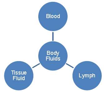
Body Fluids
Quick Review
➢ Circulatory system is one of the most important System of the body because it ensure the exchange of substances between cells of the body and external environment and transport them from one part to another.
➢ Body fluids are the medium of transport in the body. These fluids have the ability to pick up substances and distribute them to various parts of the body.
➢ Blood is fluid connective tissue. It is opaque, viscous and has ph 7.3 – 7.4. Blood has two components – Plasma and formed elements.
➢ Plasma is a faint yellow having 90 – 92 % water, 1 – 2 % salts, 7 – 8 % proteins, absorbed food, urea etc.
➢ Formed elements are of two types – Blood Corpuscles and Platelets.
➢ Blood Corpuscles are of two types – RBCs and WBCs
➢ RBCs or erythrocytes are the most abundant cells in the human body. The RBC is bounded by plasmalemma. It is non-nucleated and lack of endoplasmic reticulum which makes them more flexible thus, increasing the surface area to volume ratio for carrying more oxygen.
➢ RBCs are red in colour due to the presence of a red coloured pigment called haemoglobin which acts as a oxygen carrier molecule.
➢ One molecule of haemoglobin is made-up of four heme molecule and four globin molecules. One molecule of haemoglobin combines with four oxygen molecules
Hb4 + 4O2 → Hb4O8
➢ The average life-span of RBC is 120 days, after which it gets destroyed in the spleen.
➢ Main function of RBC is to transport O2 and CO2.
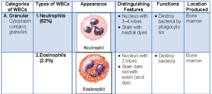
➢ WBCs or Leucocytes are most active and motile constituent of blood. They differ from RBCs as they are nucleated and lack of coloured pigment haemoglobin.
➢ WBCs are irregular in shape and are generally short lived i.e only for 12-14 days. They are of the following two types – Granulocytes and Agranulocytes.
➢ Granulocytes are WBC with granules in cytoplasm. These are further divided into three types –
(a) Basophils (b) Eosinophils (c) Neutrophils
(a) Basophils are 0.5 – 1% of WBCs and are stained by basic dyes. They produce anticoagulant.
(b) Eosinophils are 1.5% of total WBCs are stained by acidic dyes. They neutralize the toxic substances produced by pathogens.
(c) Neutrophils are 70% of WBCs and are equally stained by acidic and basic dyes. The neutrophils squeeze out from blood capillaries and fight with the foreign bacteria.
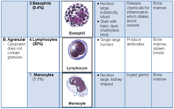
➢ Agranulocytes are the WBCs without granules in the cytoplasm.
These are two types-
(a) Monocytes (b) Lymphocytes
(a) Monocytes engulf bacteria thus they are for defence.
(b) Lymphocytes are 20-35% of WBCs. These secretes antibodies.
➢ Immunity: WBC’s protect our body from infections – where any foreign particles or pathogen enters in the body’s WBC’s like neutrophils & manacles can phagocytose them. Also, WBC’s can squeeze out through the capillary wall by the process caused diapedesis. The WBC’s engulf & destroy foreign particles. They also produce antibodies against the foreign particles (antigen). Hence, they are also called “Soldiers of the body.”
➢ Thrombocytes or blood platelets are minute oval or round, enucleated structure found floating in the blood. Their life-span is 3-5 days and get destroyed in the spleen along with RBC. The main function is to help in clotting or coagulation of blood.
➢ Extracellular fluid or tissue fluid is a solution which accumulates in the intercellular spaces. During blood flows through the capillaries, plasma and leucocytes from the blood gets leaked out through their walls and form a tissue fluid.
➢ Tissue fluid acts as a fuelling station in terms of cell nutrients and it contains glucose, fatty acids, salts and minerals such as calcium, potassium and magnesium.
➢ Lymph is the fluid along with some WBCs in intercellular spaces. Lymph acts as a middle man between blood and cells of tissues.
TOPIC-2
Blood Clotting, Blood Groups and Blood Transfusion
Quick Review
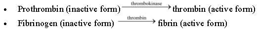
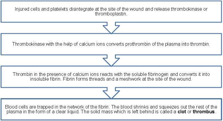
➢ Blood clotting is the natural device to check bleeding. Normal blood clotting time is 4-10 minutes.
➢ Blood platelets accumulates at the site of injured tissue cells release thromboplastins or factor ‘X’. Then thromboplastin with the help of Ca2+ convert inactive form prothrombin to active form thrombin. Thrombin acts as enzyme (Prothrombinase) along with Ca++ reacts with soluble fibrinogen and convert it into soluble form or fibrine, a solid substance that forms threads and finally form a clot.
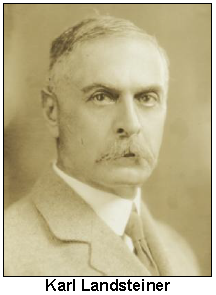
➢ Landsteiner (1900) discovered three blood groups in human, for which he was awarded by nobel prize.
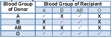
➢ The main blood groups are A, B, AB and O. This grouping is mainly based on the type of antigen and antibodies present on the surface of red blood corpuscles (RBC). The representation of different antigens and antibodies are depicted in the table given below :
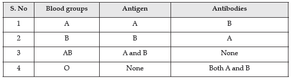
➢ Blood of group A may be transfused to persons with Blood group A and AB.
➢ Blood of group B may be transfused to persons with Blood group B and AB.
➢ Blood of AB group may be transfused to persons with Blood group AB only.
➢ Blood of O group may be transfused to persons with Blood group A, B, AB and O.
➢ O blood group can donate blood to all and hence is termed universal donor. AB Blood group persons can receive blood from all persons and hence are universal recipients.
➢ Landsteiner (1940) discovered a protein in the RBC of Rhesus monkey. Later it was discovered in some human beings also. This protein was named as Rh factor Rh antigen.
➢ Persons having Rh antigen are described as Rh+ and those without this are described as Rh–. 93% of Indian people are Rh+ and 7% are Rh–.
➢ If Rh+ blood is transfused to Rh– person, agglutination takes place. First transfusion is not serious, repeated transfusion with brief gap cause death of the recipient.
➢ Incompatibility during Pregnancy : A serious problem arises if an Rh–ve foetus. The Rh+ve blood of the foetus will stimulate the formation of anti Rh factors or antibodies in the mother’s blood. During the first pregnancy, enough antibodies are not produced to harm the foetus. During the second pregnancy, if the foetus is Rh+ve more antibodies will be produced in mother’s blood, a large number of RBC’s of the foetus are destroyed. This causes death of the foetus. This is called erythroblastosis fetalis.
TOPIC-3
Circulatory System
Quick Review
➢ Circulatory system in human beings comprised of a heart and blood vessels i.e. arteries, veins and capillaries. In different organisms, circulatory system is of two types – open and closed circulatory system.
➢ In open circulatory system, blood pumped from the heart through blood vessels but then it leaves the blood vessels and enters into body cavity e.g. arthropods, molluscs etc.
➢ In closed circulatory, the flow of blood occurs inside the blood vessels and contain a respiratory pigment haemoglobin e.g. human, birds etc.
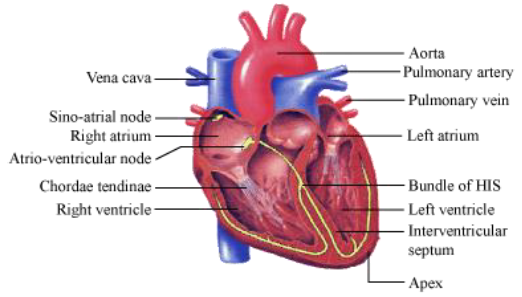
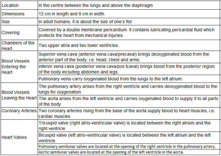
➢ Heart is the main pumping organ located in the thoracic cavity in human beings. It is enclosed in a double layered pericardium. The two layers of pericardium are separated by a narrow pericardial cavity and is filled with pericardial fluid. This fluid protects the heart from external shock.
➢ Heart is four chambered structure in human beings.
➢ Auricles – There are two auricles in the mammalian heart ( left and right ). They are separated from each other by inter auricular septum.
➢ Right auricle receive venous blood from different parts of the body ( except lungs ) through three major veins. These are two precaval veins and one post caval.
➢ Left auricle receive oxygenated blood from lungs. It is collected by pair of pulmonary veins.
➢ Ventricles – There are two thick walled muscular ventricles ( left and right ) in mammalian heart. Left ventricles is wider than right one. Auricles and ventricles are separated by an auriculo ventricular septum. The two ventricles are separated from each other by an inter ventricular septum. Right auricle opens into right ventricle through right auriculo ventricular aperture. It is guarded by a tricuspid valve. It allow the blood to flow into right ventricle only.
➢ Left auricle opens into left ventricle through left auriculo ventricular aperture. It is guarded by bicuspid valve or mitral valve. It allow the blood to flow into left ventricle only.
➢ From the right ventricle arises the pulmonary aorta which goes to lungs and carry deoxygenated blood. The opening of pulmonary aorta is guarded by three semilunar valves.
➢ From the left ventricle, left systemic aorta arises and supply oxygenated blood to all parts of the body. The opening between left ventricle and systemic aorta is guarded by three semilunar valves. They allow the blood to flow into systemic aorta only.
➢ Functioning of heart : Heart acts as a force pump and also as a suction pump. Contraction of heart is known as systole and relaxation is known as diastole. A systole and its following diastole constitute a heart beat.
➢ Relation between the size of the body & rate of heart beat of an organism → smaller the size, faster the heart rate. This is because smaller the animal, the more it loses body heat due to higher surface volume ratio, & therefore, increased heart rate distributes body heat faster. Human body, have higher metabolism for body growth & therefore, the faster heart rate keeps the ‘supply’ & ‘take off’ of the metabolic substance in right quantity.
➢ The place where contraction is originated is called pace maker. It is formed by modified cardiac muscles. Hence it is called myogenic pace maker. Heart having myogenic pace maker is called myogenic heart.
➢ In mammalian heart, two pace makers are present, viz., sinu auricular node or SA node ( in the wall of right auricle) and auriculo ventricular node or AV node ( also present in the lower side of right auricle which strengthen the signal generated by SA node.
➢ Right auricle receive impure blood from various parts of the body through two precaval veins and one post caval vein and at the same time left auricle receive pure blood from lungs through pulmonary arteries.
➢ Then systole originates in S.A node and spreads in the wall of auricles. Thus they contract and blood flows into respective ventricles.
➢ The wave of contraction is received by A.V node. It amplify the intensity and pass the wave of contraction into the walls of ventricles. The ventricles contract, pure blood from left ventricle into systemic aorta and impure blood from right ventricle flows into pulmonary aorta.
➢ Sounds created by the valves of heart during its contraction are called heart sounds. When auricles relax, the bicuspid and tricuspid valves snap shut. It creates the first sound Lubb. When ventricles relax, the semilunar valves are closed. It creates the second sound called Dubb.
➢ Cardiac cycle is the sequence of the events that takes place in heart during one heart beat. Its duration is 0.8 seconds.
➢ Blood vessels are the branched tubes which extends from heart to all parts of the body. There are three main types of blood vessels which carry blood vessels towards and away from the heart. These are arteries, veins and capillaries.
➢ Artery is a blood vessel whose function is to carry blood away from the heart and towards any other organs. An artery comprised of thick walls and narrow central lumen.
➢ Vein is a type of blood vessel which carry blood away from the organs and towards the heart. It is comprised of thinner walls and a large central lumen then arteries.
➢ Capillary is a narrow tube like blood vessel, comprised of single layer of endothelial cells. Capillaries have the ability of contracting and dilating with the decrease and increase in the blood supply to various body parts.
➢ Blood in human beings flows twice in the heart before it completes one round. It is comprised of one short flow, i.e. pulmonary circulation and long flow, i.e. systemic circulation. Due to this reason the blood flow in human beings is known as double circulation.
➢ In pulmonary circulation, deoxygenated blood is collected from the heart ( right ventricle ) by a pulmonary artery and returns oxygenated blood to the heart ( left auricle ) through pulmonary veins.
➢ In systemic circulation, oxygenated blood from the heart, ( left ventricle ), is carried to different parts of the body through aorta that carries deoxygenated blood into the heart ( right auricle ) through vena cava.
➢ Hepatic portal circulation consists of a hepatic portal vein that returns the blood from intestine and breaks into capillaries in liver. Thus, the liver cells can take up nutrients brought by it from the small intestine. In liver these excess of nutrients are stored and allow remaining required amount of nutrients to flow in the blood.
➢ Hepatic artery and hepatic portal vein are the blood vessels that enter the liver. Hepatic vein leaves the liver. Renal artery enters the kidney and renal vein leaves the kidney.
➢ Pulse is the wave of raised blood pressure due to pumping activity of the left ventricle . It is equal to heart beat and is felt extremely in the radial artery near the wrist.
➢ Blood pressure is the force of blood against the walls of blood vessels. The rise of blood pressure during contraction of heart is called systolic pressure and the fall of blood pressure during relaxation of heart is known as diastolic pressure. The normal blood pressure in adults is 100-140 mm Hg (systolic pressure) and 60-80 (diastolic pressure). A rise in blood pressure is called hypertension and decrease in blood pressure is called hypotension.
Know the Terms
➢ Haemocoel : It is cavity contained blood.
➢ Pulse rate : It is the difference between systolic B.P and diastolic B.P. and it is 40 mm Hg.
➢ Fossa ovalis : It is an oval depression present in interauricular septum at point where an opening called foramen ovale is present in embryo.
➢ Artificial pace maker : It is an artificial electronic device which regularly sends small amount of electric charge for maintaining rhythmicity of heart.
➢ Atherosclerosis : It is due to loss of elasticity of coronary arteries, which increases blood pressure.
➢ Thrombus : It is a clot of blood formed in the body.
➢ Embolus : Mobile thrombus is called embolus.
➢ Electrocardiogram (ECG) : It is the recording of electrical events which take place by the spread of cardiac impulse.
➢ Electrocardiograph : A machine used for electrocardiography.
➢ Cardiac centres : Group of neurons in the floor of medulla which controls rate of the heart beat.
➢ Chordae tendineae : White fibrous threads which extend from bicuspid and tricuspid valves to the papillary muscles of ventricles.
➢ Coronary circulation : Circulation of blood to and from the wall of heart.
➢ Lymph nodes : The masses of lymphoid tissue which produce agranular WBCs.
➢ Mean arterial pressure : The blood pressure determining the average rate of blood flow inside the blood vessels.
➢ Murmur : Abnormalities in the heart-sounds due to defective heart valves.
➢ Neurogenic heart : When the contraction of heart is initiated by the nerve ganglion present near the heart and send nerve impulses. It is found in arthropods and molluscs.
➢ Papillary muscles : These are large sized ridges on the wall of ventricles. Chordae tendineae are attached on the papillary muscles.
➢ Stroke volume : Amount of blood ejected from the heart in one heart beat. (i.e. 70ml)
Components of Blood and Lymph (Detail Study)
Need for Transport inside Our Body
• In Digestive System: The nutrients absorbed from the digested food need to be transported to each cell to perform their functions.
• In Excretory System: All the wastes generated need to be collected from whole body and flushed out.
• In Endocrine System: The hormones produced need to be sent to each and every part of our body.
• In Respiratory System: The oxygen and CO2 need to be transported through out the body.
Blood: Connective tissue consisting of fluid matrix, plasma, and formed elements
Functions of Blood
(i) Transportation
• Transport of digested food from alimentary canal to tissues
• Transport of oxygen from lungs to the tissues
• Transport of carbon dioxide from tissues to lungs
• Transport of excretory material
• Distribution of hormones from endocrine glands
• Distribution of heat throughout the body
(ii) Protection
• Formation of clot in case of cut, thus preventing blood loss
• Protecting body from bacteria
• Production of antitoxins and antibodies
Components of Blood: It consists of fluid part, called plasma, and cellular elements that consist of red blood cells, white blood cells, and platelets.
Plasma
• 55% of blood
• Plasma = 90-92 % water + 6-8% proteins
• Proteins present
Fibrinogen − blood clotting
Globulins − defence mechanisms
Albumins − osmotic balance
• Also contain mineral, glucose, amino acids, and lipids in traces Blood clotting factors are present in inactive form in plasma.
• Serum = Plasma − Clotting factors
Formed Elements

Red Blood Cells (RBCs) – These are responsible to carry oxygen through the body.
• Haemoglobin : A chief chemical constituent of RBCs. It is present inside stroma – a spongy body of RBCs.
• It is made up of iron and protein.
• It easily combines with oxygen forming oxyhaemoglobin, an unstable compound that easily donates oxygen to the needy tissues.
• It also carries a small amount of CO2 in the form of carbaminohaemoglobin.
• Carbon monoxide Poisoning
• Haemoglobin has high affinity towards carbon monoxide as it forms a more stable compound carboxyhaemoglobin (HbCO).
• It results in decreased efficiency of oxygen transport by blood, leading to less supply of oxygen in the body.
• It may result even in death.
Increased Efficiency of RBCs
The mammalian red blood cells are more efficient as compared to others as they lack certain cell organelles. The factors that makes them more efficient are:
• Loss of nucleus: This makes them biconcave in shape hence, increasing their surface area to volume ratio to maximise oxygen absorption.
• Loss of mitochondria: Lack of mitochondria means that no cellular respiration can occur in the RBCs. Thus all the oxygen absorbed from the lungs are transported to the tissues as they don’t need it for themselves any more.
• No endoplasmic reticulum: It results in increased flexibility for their movement through the constricted capillaries.

Functions of Leucocytes (WBCs)
The basic function of white blood cells is body defence.
• Phagocytosis: This is a defence mechanism in which the WBCs engulf the solid substances like bacteria.
• Inflammation: Inflammation is a result of reaction of tissues to injuries and to localised invasion of germs. The leucocytes (especially monocytes and neutrophils) reach the inflamed area by migrating through blood vessel walls (diapedesis). They can then fight against the disease causing germs and also destroy the damaged cells by phagocytosis.
• Formation of Antibodies: These are produced by WBCs (lymphocytes) to kill or neutralise the germs and poison from them. These are stimulated by introducing weakened germs through vaccination.
Lymph
• Lymph is the fluid released out of blood capillaries leaving behind larger proteins and formed elements.
• It consists of water and some water soluble substances.
• It has some mineral distribution as present in plasma.
• The network of lymph vessels composes lymphatic system.
Uses
• Lymph contains lymphocytes that are involved in immune response.
• Lymph carries nutrients, hormones, etc.
• Lymph absorbs fats in lacteals found in intestinal villi.
Blood Coagulation:
• Clotting is required to prevent excessive loss of blood from the body.
• Blood clot – formed by threads of fibrin in which formed elements are trapped.
• Mechanism of coagulation is a cascade of reactions involving several clotting factors.
• Calcium plays an important role in blood clotting mechanism.
• Serum : The Clear liquid squeezed out of the network of fabrin in which the blood cells are trapped is called Serum.
Blood Groups and Rh Factor
Blood groups
• Widely used blood grouping − ABO and Rh
ABO Grouping
• Surface antigens A and B are present on RBCs.
• Antibodies are produced against corresponding antigens.

• Universal Donor − Blood group ‘O’
• Universal recipient − Blood group ‘AB’
Rh Grouping
• Individuals with Rh antigens present on RBCs are Rh positive and those without it are Rh negative.
• If Rh −ve mother bears an Rh +ve child during first pregnancy when mother’s blood is exposed to Rh +ve antigens, then anti − Rh antibodies are produced in her blood.
• During subsequent pregnancies, these antibodies may destroy RBCs of the foetus. This results in severe anaemia and jaundice to new born. This condition is called erythroblastosis foetalis.
• During Rh incompatibility, the first child is safe or may have anaemia.
• However, this condition can be avoided for subsequent pregnancies by administering anti-Rh antibodies of mother immediately after delivery of first child.
Human Circulatory System
• Humans have a closed circulatory system: Blood pumped by the heart always flows through a closed network of blood vessels.
• Human circulatory system consists of:
• Muscular, four-chambered heart
• A network of closed, branching blood vessels − veins, arteries and capillaries
• Blood
Blood vessels
Arteries are tough, elastic tubes that carry blood from the heart and supply it to various organs of the body. As the arteries move away from the heart (i.e., on reaching organs and tissues), they divide into smaller vessels.
Arteriole is the smallest or the final branch of artery. These are highly muscular and can easily change their diameter. Arteries are red in colour because they carry oxygenated blood.
The smallest blood vessels are called capillaries. They have very thin walls and lack muscles. The capillaries can easily dilate (vasodilation) and contract (vasoconstriction), thus can regulate the blood supply to different organs.
Functions Of Capillaries:
• It allows the outward diffusion of Oxygen
• It allows the WBCs to squeeze out of capillary walls
• It allows inward and outward diffusion of urea, glucose, hormones etc.

Capillaries in organs and tissues gradually reunite and increase in size. The smallest of the united common branch are called a venule. The venules then join to form the veins. Veins collect blood from different organs and tissues and transport it to the heart.
They are thin-walled as compared to arteries. This is because they bring back blood from the organs to the heart and blood is no longer under pressure. These veins carry deoxygenated blood into the heart.
Differences between Arteries and Veins

Hepatic portal system
Hepatic portal system consists of a network of veins that facilitate the recirculation of blood to the liver from digestive tract and spleen. The major blood vessels of this system includes hepatic portal vein, inferior mesentric vein, superior mesentric vein and gastrosplenic vein.
Significance of hepatic portal system
Hepatic portal system allows metabolization of digested substances in the liver before they are propagated to the systemic circulation. Certain toxic substances can be inactivated by the liver metabolism and excreted from the body. Thus, hepatic portal system plays an important role in the elimination of toxic substances from the body.
Practical significance of hepatic portal system can be observed in the filed of pharmacology. Many drugs such as nitroglycerine can be inactivated through liver metabolism. Such drugs are therefore not administered through oral means because hepatic portal system can transfer them to the liver and make them ineffective.
Structure of Heart
Heart
• Location: Thoracic cavity in between the lungs; slightly tilted to the left
• Protected by a double-walled pericardium, enclosing the pericardial fluid
• Has 4 chambers: 2 upper chambers − right and left atria
• 2 lower chambers − right and left ventricles
• Inter-atrial septum: Separates the right and the left atria
• Inter-ventricular septum: Separates the right and the left ventricles
• Atrio-ventricular septum: Separates the atria and the ventricles of the same sides
• Septa have openings through which the two chambers on the same sides are connected.
• Tricuspid valve: Present between the right atria and the right ventricle
• Bicuspid (mitral) valve: Present between the left atria and the left ventricle
• Semilunar valves: Guard the openings of the right and the left ventricles into the pulmonary artery and the aorta respectively.
• Special cardiac musculature called nodal tissue is distributed throughout the heart.
• Sinoatrial node (SAN): Present at the upper right corner of the right atrium
• Atrio-ventricular node (AVN): Present at the lower left corner of the right atrium
• AV bundle (a bundle of nodal fibres) continues from the AVN and passes through the atrio-ventricular septa to reach the inter-ventricular septum.
• There, it divides immediately into right and left bundles. From these branches, minute fibres arise throughout the ventricular musculature. These fibres are called purkinje fibres.
• Right and left bundles + Purkinje fibres = Bundle of His
• Significance of nodal musculature: Auto-excitable; generates and maintains action potential to sustain the rhythmic contraction activity of the heart
• Pacemaker of the heart − Sino-atrial node (SAN)
• Heart beats 70−75 times/min
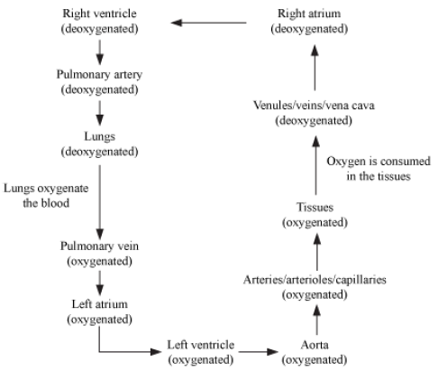
Double Circulation and Cardiac Cycle
Double Circulation
• In human beings, oxygenated blood is received by the left atria while deoxygenated blood is received by the right atria, which then pass it on to their respective ventricles.
• This prevents the oxygenated and deoxygenated blood from mixing. This unique pathway is called double circulation.
• Double circulation consists of two parts: pulmonary circulation and systemic circulation.
• In systemic circulation, the deoxygenated blood is collected from all the body parts and transported to the heart through vains. The collected blood is poured into the right atrium through superior and inferior vena cava. Once the blood is oxygenated, it is transported back to various body parts from the left ventricle of the heart through aorta.
• In pulmonary circulation, pulmonary artery collects deoxygenated blood from the right ventricle of heart and carries it to the lungs. After gaseous exchange in the blood, the pulmonary veins collect the oxygenated blood from the lungs and carry it to the left atrium of the heart.
Blood Circulation Pathway
Hepatic Portal System
• Portal Vein : A vein that starts and ends with capillaries.
The veins coming from intestines and stomach does not directly delivers the blood to the posterior vena cava. Instead, they enter into the liver combined together as hepatic portal vein, which then splits into numerous capillaries. This is opposite to the characteristic of vein. Later these capillaries combine to form hepatic vein which later joins the posterior vena cava. This whole system is known as hepatic portal system.
• Hepatic portal system helps in assimilation of different nutrients absorbed by blood during digestion process in the liver.
• It also helps in detoxification of blood in the liver.
Cardiac Cycle
• Cardiac cycle is the sequence of events which occur from the beginning of one heart beat to the beginning of the next heart beat.
• In the beginning, all the 4 chambers of the heart are in a state of joint diastole (relaxation).
• Tricuspid and bicuspid valves open and blood from the veins and the vena cava flow into the atria, and then into the ventricles because of the opening of the valves.
• SAN generates an action potential, and both atria undergo contraction (Atrial systole).
• The flow of blood into the ventricles increases by 30%.
• The action potential is conducted towards the ventricles through the AVN and the AV bundles, from where the bundle of His transmits this action potential over the entire cardiac musculature.
• The ventricles contract (ventricular systole) and the atria relax (atrial diastole) as a result of the conduction of action potential.
• Ventricular pressure increases. Hence, bicuspid and tricuspid valves close, to prevent the backflow of blood into the atria. Further increase in pressure in the ventricles leads to the opening of the semilunar valves.
• Blood from the ventricles flow into the pulmonary artery and the aorta, and subsequently into the circulatory pathways.
• Consequently, the ventricles relax (ventricular diastole), ventricular pressure falls, and the semilunar valves close to prevent the backflow of blood into the ventricles.
• Ventricular pressure further falls. As a result, the bicuspid and tricuspid valves open. This is because pressure is exerted on the atria by the blood entering them through the veins.
• Once again, joint diastole is experienced and the entire cycle is repeated.
Pulse
The distension felt because of the contraction of heart, every time when blood passes through the arteries, is referred as pulse. This alternate expansion and recoil of the arteries occur because of the elastic nature of artery walls. A pulse rate can give indirect measure of the heart beats.
Blood Pressure
• The pressure exerted by blood through the arteries on their walls.
• There are two limits to the blood pressure:
• Systolic Pressure (upper limit): When fresh blood is pushed through artery due to ventricular contraction of heart
• Diastolic Pressure (lower limit): When the wave has passed over
• The normal blood pressure for an adult is 120 (systolic) and 80 (diastolic)
Cardiac output
• Heart beats: Average 72 times/minute (heart rate) • Duration of cardiac cycle is 0.8 seconds.
• Stroke volume: Amount of blood pumped by the heart in one cardiac cycle Stroke volume = 70 mL
• Cardiac output = Stroke volume × Heart rate
= 70 mL × 72 times / min
~ 5000 mL
Heart Sounds
• Lub: First heart sound, associated with the closure of the tricuspid and bicuspid valves
• Dub: Second heart sound, associated with the closure of the semilunar valves
• These heart sounds are of diagnostic significance.
Every organ in our body requires the involvement of the circulating body fluids.
Blood
Blood is a never-stationary fluid and it is always in motion from the heart to the arteries and back through the veins.

Functions of Blood
• Blood forms a clot which serves to prevent the loss of blood and the entry of disease-causing germs.
• White blood cells protect the body from diseases by engulfing bacteria which may have entered the body.
• Antibodies produced by the blood neutralise poisonous substances or kill germs which enter the body.
• Blood transports digested food from the alimentary canal to the tissues.
• It transports excretory materials from the tissues to the liver, kidneys or skin for elimination.
• Blood helps in keeping the temperature of the body uniform by distributing heat.
• Haemoglobin of RBCs combines with oxygen to form oxyhaemoglobin which reaches tissues to deliver the oxygen.
Composition of Blood
Blood is made up of plasma and the blood corpuscles
Plasma
It is a light yellow – coloured alkaline liquid.
It mainly consists of

Cellular Elements
There are three kinds of cellular elements found in the blood:

Haemoglobin
Haemoglobin is a respiratory pigment present in the stroma of RBCs. It combines readily with oxygen to form an unstable compound oxyhaemoglobin. This compound delivers oxygen to tissues.
Haemoglobin has a very strong affinity for carbon monoxide. When combined with carbon monoxide, it forms a stable compound carboxyhaemoglobin.
Carboxyhaemoglobin reduces the capacity of the blood in transporting oxygen, sometimes even resulting in death.

Different types of White Blood Cells

Clotting of Blood (Coagulation)
Blood Transfusion
• Sometimes, it is necessary to inject blood into the body of patients undergoing surgery. This is called blood transfusion.
• The German biochemist Karl Landsteiner was the first to suggest that the blood of different individuals vary.
There are several systems of blood grouping. The ABO system and the Rh system are the most important.
ABO System
• According to the ABO blood group system, there are four blood groups A, B, AB and O.
• O type blood can be given to persons of all types of blood, i.e. O, A, B and AB. Hence, a person with O type of blood is called a universal donor.
• A person with AB type of blood can receive blood from all types, i.e. AB, A, B and O. Hence, such a person is called a universal recipient.
Compatibility and Incompatibility in the ABO System
Rh System
• The blood of most people contains a substance called Rh factor.
• Rh stands for Rhesus, our common primate ancestor in which this factor was first discovered.
• When the blood of an Rh positive (Rh+) individual is transfused into a person lacking the Rh factor, the blood of the recipient develops antibodies against the Rh factor which may even lead to death
Tissue Fluid (Intercellular Fluid)
• As blood flows in the capillaries of tissues, the plasma of leucocytes leaks out through their walls and bathes the cells. This fluid is called tissue fluid or intercellular fluid.
• Cells absorb oxygen and other nutrients and give out carbon dioxide to the tissue fluid.
Lymph and Lymphatic System
• Most of the tissue fluid enters another set of vessels called lymphatic vessels, and this fluid is called lymph.
• Lymph vessels drain lymph into lymph nodes.
• From lymph nodes, through lymph vessels again, lymph enters the vena cava just before its entry into the right auricle

Function of Lymph
• Supplies nutrition and oxygen to parts where blood cannot reach.
• Drains away excess tissue fluid and metabolites.
• Lymphocytes and monocytes of the lymph help in the defense mechanism of the body.
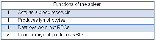
The Spleen
• The spleen is a large lymphatic organ, about the size of a clenched fist.
• It is reddish brown in colour and situated in the abdomen behind the stomach and above the left kidney
The Circulatory System
• Blood in our body circulates in a closed manner, i.e. through blood vessels, all the time. Such type of blood circulation is called a closed vascular system.
• In animals such as insects, the blood mostly flows through open spaces, and such type of circulation is called open vascular system.
• The human blood circulatory system consists of heart, arteries, veins and blood capillaries
The Heart


Circulation of Blood in the Heart
The circulation of blood in the heart occurs due to alternate contraction and relaxation of the heart chambers. Contraction is also known as systole, while relaxation is also known as diastole. The series of events which occur during one complete beat of the heart is called cardiac cycle


• Cardiac muscles contract rhythmically in response to self-generated impulses.
• The pacemaker of the sino-atrial node (SA node) is located in the upper wall of the right atrium. It triggers an impulse which causes an atrial systole.
• This impulse quickly reaches the atrio-ventricular node (AV node) located at the bottom of the right atrium which initiates a ventricular systole.

The rate of the heart beat varies among different species. Smaller the size of the animal, faster is the heart rate
Blood Vessels
The blood vessels are branched tubes extending from the heart to all parts of the body.
An artery is a vessel which carries blood away from the heart towards any organ.
A vein is a vessel which carries blood away from an organ towards the heart.
A capillary is a very narrow tube of about 8 μm in diameter.
Pulmonary and Systemic Circulation

Pulmonary and Systemic Circulation
Pulmonary circulation pertains to the lungs. It starts in the pulmonary artery. It sends the deoxygenated blood to the lungs. Pulmonary veins collect oxygenated blood from the lungs and carry it back to the heart.
Systemic circulation pertains to the major circulation of the body. The aorta receives blood from the heart and sends it to the various parts of the body. Veins collect the deoxygenated blood from body parts and pour it back into the heart.
Hepatic Portal System

• Before conveying the blood to the posterior vena cava, the veins of the stomach and intestine enter the liver as a combined hepatic portal vein.
• The hepatic portal vein divides into capillaries and then forms a new hepatic vein. Portal Vein: It is a vein which starts with capillaries and ends in capillaries.
• In the liver, excess nutrients are stored, toxic substances are detoxified and excess amino acids are broken down.
Pulse
• The pulse is the alternate expansion and elastic recoil of the wall of the artery during ventricular systole.
Blood Pressure
• Blood pressure is the pressure which the blood flowing through the arteries exerts on their walls.
• There are two kinds of blood pressure:
• Systolic Pressure: The upper limit of the pressure. It occurs each time when the heart contracts and fresh blood is pumped into arteries.
• Diastolic Pressure: The lower limit of the pressure. It occurs each time when the heart is in diastole, i.e. this pressure is observed between two heart beats.
• The normal blood pressure of an adult human is 100–140 mm (systolic) and 60–80 mm (diastolic).
• A sphygmomanometer is an instrument used to measure blood pressure