Students should refer to Nervous System ICSE Class 10 Biology notes provided below designed based on the latest syllabus and examination pattern issued by ICSE. These revision notes are really useful and will help you to learn all the important and difficult topics. These notes will also be very useful if you use them to revise just before your Biology Exams. Refer to more ICSE Class 10 Biology Notes for better preparation.
ICSE Class 10 Biology Nervous System Revision Notes
Students can refer to the quick revision notes prepared for Chapter Nervous System in Class 10 ICSE. These notes will be really helpful for the students giving the Biology exam in ICSE Class 10. Our teachers have prepared these concept notes based on the latest ICSE syllabus and ICSE books issued for the current academic year. Please refer to Chapter wise notes for ICSE Class 10 Biology provided on our website.
Nervous System ICSE Class 10 Biology
TOPIC-1
Structure of a Neuron
Quick Review
➢ Living organisms have the ability to respond and react to their surrounding environment. Plant responses are regulated by chemical substances called phytohormone. Animals posses nervous system and hormones for control and coordination of various functions.
➢ Nervous system of animals are formed by nervous tissue, which is composed of neuron and neuroglia cells.
➢ A neuron is a structural and functional unit of nervous system. Neurons are the longest cells in the body.
➢ A neuron has three components – Cell body, Dendrites and Axon.
➢ A cell body is like a typical cell containing nucleus and granular cytoplasm. Dendrites are short and branched cytoplasmic protection. Axon is a single, elongated fibre. Both axon and dendrites arise from cell body.
➢ Axon is covered by myelin sheath. The gaps between the two adjacent myelin sheath are called nodes of Ranvier. The terminal portions of the axons have swollen bulb-like structure called synaptic knob which store certain chemicals called neurotransmitter. The axon transmit impulses away from the cell body to a synapse or to a neuromuscular junction.
➢ When a neuron is suitably stimulated, an electrical disturbance is generated which swiftly travel along its plasma members, arrival of the disturbance at the Neuron endings or out put zone. Triggers events that may cause stimulation or inhibition of adjacent neurons & other cells the terminal to the dendrites of another neurons to carry impulses from one neuron to another Neuron . This close proximity is called synapse.
➢ During impulse transmission, synapse is filled with acetylcholine. It is impulse specific. This chemical diffuses across the synaptic cleft and attaches to receptor site on the dendrites of next neuron. There it causes the depolarization of the membrane and initiates new impulse.
➢ Immediately after impulse transmission, acetylcholine is hydrolysed by an enzyme, choline esterase. This enzyme splits the acetylcholine into acetic acid and choline. This prevents the continuous stimulation of dendrite. The acetic acid and the choline return by diffusion into synaptic cleft where these are again recombined with the help of synthesizing enzymes into acetylcholine.
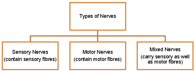
➢ Neurons are of three types – Sensory neurons, Motor neurons and Mixed neurons.
➢ Sensory neurons transmit impulses towards central nervous system. Motor neurons transmit impulses from central nervous system to effector organs. Mixed neurons acts as sensory as well as motor neurons.
TOPIC-2
Human Nervous System
Quick Review
➢ The nervous system in human can be divided into two major parts :
1.Central nervous system (CNS)
2.Peripheral nervous system (PNS) :
(a) Somatic nervous system. (b) Autonomic nervous system.
➢ The central nervous system consists of brain and spinal cord.
➢ Brain of man weighs about 1.4 kg. It is well developed and lodged and protected in a bony box, called cranium.
➢ In the cranium, brain is covered by three layers, called meninges. These are – Outer duramater, Middle arachnoid layer, Inner piamater.
➢ Duramater and arachnoid layer separated by sub dural space. Arachnoid layer and piamater are separated by sub arachnoid space.
➢ Sub dural space and sub arachnoid space are filled with cerebrospinal fluid (alkaline in nature). It protects the brain from shocks, mechanical injuries and act as middle man between nervous tissue and blood.
➢ Brain is formed by two types of substances – Inner gray matter (contain cell bodies of neurons) and outer white matter (contain proximal parts of axons).
➢ Human brain is divisible into three main parts – Forebrain, Midbrain and Hindbrain.
➢ Forebrain is the largest part of the brain and is divided into olfactory lobes, cerebrum and diencephalon.
➢ Olfactory lobes are paired lobes and form the anterior most part of the forebrain. Each olfactory lobe has an olfactory bulb and an olfactory duct. These lobes are functionally related to smell.
➢ The cerebrum is the largest and most prominent part of the brain. It is divided into right and left hemispheres. Cerebral hemisphere is hollow from the interior and has two regions i.e. outer cortex and inner medulla.
➢ Each hemisphere is divided by gyri and sulci into distinct areas known as frontal, parietal, temporal and occipital lobes. The cerebral cortex is the highest centre for many activities. There are many functional areas of the cerebrum which include visual area, auditory area, association area.
➢ Diencephalon consists of two major parts – thalamus (a major coordinating centre for sensory and motor signaling) and hypothalamus (which mainly controls the body temperature, urge for eating, drinking and action of pituitary gland).
➢ Mid brain is located in between the thalamus and hypothalamus and pons. It is a smaller part of the brain which help to relay information for vision and hearing.
➢ Hind brain is divided into three main parts – Cerebellum, Pons varolli and Medulla oblongata.
➢ Cerebellum is well developed and is composed of a median lobe called vermis and a pair of lateral lobes called flocculi. Grey matter of vermis is branched and is known as arbor vitae. It is concerned with maintaining equilibrium.
➢ Pons varolli is located below the cerebellum and is responsible for carrying impulses from cerebellum to cerebrum. It also helps in regulation of breathing movements.
➢ Medulla oblongata is the posterior most part of the brain and is located underneath the cerebellum. It is the most essential part of brain, therefore, it controls beating of heart, movement of alimentary canal, movements of lungs, movement of diaphragm etc.
➢ Spinal cord is a tube like, present in the nature canal of vertebral column. It is covered by three meninges piamatter, duramatter and arachnoid matter. In the centre is present centre canal. Grey matter forms a central portion and resembles the alphabet H. White matter forms the outer portion and is composed of medullated nerve fibres. The bundle of nerve fibres ascend and descend along the white matter and are called as nerve tracts.
➢ Spinal cord controls reflex actions of the body. It conducts sensory and motor impulses to and from the brain.
➢ Peripheral nervous system is the lateral or side part of nervous system which connects central nervous system with different parts of body with receptors and effectors i.e. sense organs and muscular system of the body, e.g. cranial nerves and spinal nerves.
➢ Nerves formed from spinal cord are known as spinal nerves. Each spinal nerve has two roots, viz., dorsal root and ventral root. These are fused in the neural canal and form a spinal nerve and comes out from the vertebral column.
➢ Cranial nerves are those nerves which arise from the brain. Human has 12 pairs of cranial nerves. These may be Afferent nerves, Efferent nerves and Mixed nerves and there are 31 pairs of spinal nerves in humans.
➢ Autonomic nervous system (or involuntary nervous system) controls various functions which are carried independently in the body such as control of rate of heart beat, the movement of alimentary canal etc.
➢The autonomic nervous system consist of two Antagonistic Systems (i) Sympathetic Nervous System and (ii) Parasympathetic Nervous System.
➢ Reflex action is the spontaneous automatic response to a stimulus without the will of an animal. Most of the reflex actions are controlled by spinal cord.
➢ The route through which the impulses travelled to complete a reflex action is called reflex arc. The sensory stimulus reach the dorsal half of spinal cord through dorsal root of a spinal nerve (afferent nerve). The impulse is processed and necessary stimulus was given to the efferent nerve through ventral root via intermediate neuron (present in spinal cord).

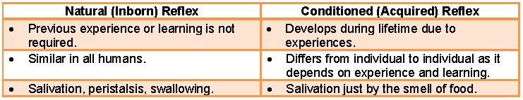
➢ Reflexes are of two types : Conditional reflexes and Unconditional (Natural) reflexes.
➢ Conditional reflexes are acquired reflexes during lifetime of an individual. They are not constant. It includes receptor, sensory nerves, area of cerebral cortex, motor nerve, effector organ. Functions of conditional reflexes – Our habits are formed by conditional reflexes, elimination of harmful influences and Ensure adaptation of the organism.
➢ Unconditional reflexes are inborn and transmitted from generation to generation e.g. Breast feeding, constriction of pupils if bright light falls on the eye.
➢ Importance of reflex action : It enables the organism for an immediate response to a harmful stimulus, It reduces the overloading in brain and increases the chances of survival of an organism.
TOPIC-3
Sense Organs – Structure and Functions of the Eye and Ear and their Various Parts
Quick Review
➢ Sense organs are the organs that enable us to detect all types of changes that occurs in the environment. These organs send appropriate signals to the central nervous system, where all the inputs are processed and analysed. The major sense organ includes eye, ear, tongue, nose and skin. These organs are responsible for vision, balance and hearing, taste, smell and feel respectively.
➢ The organ of sight and vision are a pair of eyes in humans. These are hollow, spherical organs situated in body cavities called orbits or eye sockets.
➢ The accessory structure of the eye include eyebrows, eyelids and tear glands (lacrimal glands).
➢ Eyebrows does not constitute the internal part of the eye they instead act as the protective covering and protect the eye from sweat, dust and other foreign bodies.
➢ The eyelids are two moveable folds situated above and below the front of the eye which prevents falling of larger particles into the eyes.
➢ Tear glands (lacrimal glands) are located at the upper sideward portion of the orbit and secrete tears. The tear help in keeping front surface of the eye clean by washing away the dust particles.
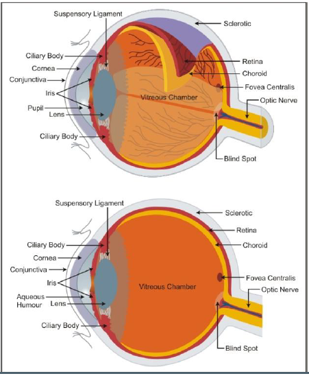
➢ Eye ball is made up of three layers – sclerotic, choroid layer and retina.
➢ The outer covering of eye is the sclerotic coat, made up of dense connective tissue. In front, it is transparent and form cornea. Cornea is covered by transparent epidermal layer, called conjunctiva. Conjunctiva is continuous with the lining of eye lids.
➢ The middle layer of eye is the choroid coat, which is highly vascular and contains pigment cells. In front, choroid coat forms the iris, which is perforated by a round pupil. Iris is visible through cornea is pigmented (black, brown, blue, green, grey etc.) in human and in other mammals.
➢ In the peripheral margin, choroid coat form a ciliary body. Behind the iris, is a biconvex lens.
➢ The inner most layer of eye is the retina, which is light sensitive. It consists of two layers – Outer layer is closely attached to the choroid coat and inner layer is light sensitive.
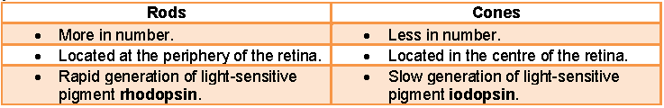
➢ Retina proper contain rods and cones. Rods contain rhodopsin. Rods distinguish the intensity of light. Cones contain iodopsin. Cones are responsible for colour distinction.
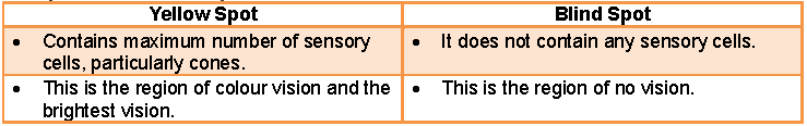
➢ Nerve cell bodies of neurons lie in retina proper. Their axons formed into optic nerve. The point of exit of optic nerve from retina forms blind spot.
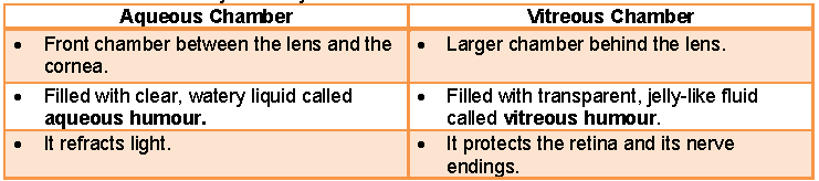
➢ Lens divided the cavity of eye ball into two chambers – (i) Anterior chamber is called aqueous chamber and is filled with aqueous humour (ii) Posterior chamber is called vitreous chamber and is filled with vitreous humour.
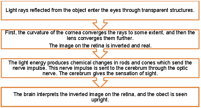
➢ Cornea place the image on the retina. Lens is adjusted to form clear image. The adjustment of lens is called accommodation. Thus an inverted image is formed on the retina. This image is interpreted in the brain and thus animal see the object in upright way.
➢ Disorders of eye – Myopia (Short sight – Corrected by using concave lens), Hypermetropia (Long sight – Corrected by using convex lens), Cataract (Lens becomes opaque and corrected by surgical transplantation of lens), Astigmatism (Caused due to irregular curvature of cornea and corrected by cylindrical lens),
➢ Human ear is concerned with two sensory functions such as hearing and maintenance of body balance. The ears are located on both sides of the head.
➢ In mammals, ear consists of three parts – external ear, middle ear and internal ear.
➢ External ear is also called Pinna. It is formed by elastic cartilage. Pinna is movable in mammals with the auricular muscles present in it. But in human being it is immovable.
➢ Opening of ear leads into tubular passage, known as external auditory meatus. Inner part of it contain ceruminous glands. They secrete ear wax that lubricate the tympanum (ear drum).
➢ Middle ear is in tympanic cavity. It is filled with air. This cavity is connected to the pharynx through eustachian tube (for equalizing air pressure on both sides of ear drum). Tympanic cavity starts from ear drum (tympanum).
➢ Tympanic cavity internally opens into the auditory capsule through a pair of windows – upper fenestra and fenestra rotunda.
➢ Behind the ear drum, middle ear contains a chain of three bones viz; malleus (hammer like), incus (anvil like) and stapes (stirrup like). They convey the vibrations of ear drum to the internal ear.
➢ Internal ear is a delicate structure, composed of membranous labyrinth. It is lodged and protected in the auditory capsule, formed by periotic bone.
➢ Space between membranous labyrinth and bony capsule is filled with perilymph. Membranous labyrinth is filled with endolymph. It contain small calcareous pieces, called otoliths.
➢ Membranous labyrinth is formed by a dorsal utriculus and ventral sacculus.
➢ Utriculus is provided with three semicircular canals (external, anterior and posterior). They are arranged at right angle to each other. Each semicircular canal ends with a bulb like structure, called ampulla. It contains sensory area known as crista ampullaris.
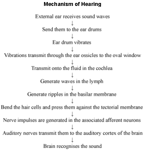
➢ Sacculus is the ventral part of membranous labyrinth. It has a spirally coiled tube, called cochlear duct or lagina membrane. This causes the tympanic membrane to vibrate.
➢ The vibrations are picked up by the malleus on the other side. These vibrations are transmitted to the fenestra ovalis via incus and stapes.
➢ These vibrations travel along the vestibular canal to the end of the cochlea. Due to this, the liquid in the cochlea begins to vibrate and the pressure vibrations are turned into electrical signals by the cochlea. These electrical signals are carried by auditory nerve to the brain.
➢ The equilibrium is maintained by semicircular canals. The equilibrium is of two types – Dynamic equilibrium and Static equilibrium.
➢ Dynamic equilibrium is maintained by cristae present in ampullae of semicircular canal . When the head is rotating, at that time sensory hair get disturbed by moving endolymph. The disturbances are conducted to sensory cells and then to nerve fibres which transmit impulses to brain. It brings the organism in equilibrium, while the similar sensory cells located in two parts of semicircular canal, i.e. utriculus and sacculus are concerned with the static equilibrium of the body, i.e. when the body is stationary.
Know the Terms
➢ Action potential : It is the potential change that occurs in an axon on stimulation of a nerve fibre.
➢ Binocular vision : Simultaneous focusing of two eyes on same object.
➢ Blind spot : The spot on the retina which has no photoreceptor.
➢ Central canal : The cavity of spinal cord.
➢ Chiasma : A cross of two optic nerves.
➢ Depolarization : The reversed polarity of the nerve fibre.
➢ Foramen of Monro : An aperture which connects lateral ventricles of cerebral hemispheres with 3rd ventricle of diencephalon.
➢ Saltatory conduction : Conduction of nerve impulse by myelinated nerve fibre on which impulse jumps from one node of Ranvier to another.
➢ Resting potential : Potential that exists in an axon at rest or without stimulation.
➢ Organ of Corti : It is hearing apparatus present in the middle canal of cochlea.
➢ Perception : It is the conscious awareness and interpretation of sensations.
➢ Neuroendocrine system : It is a network of endocrine glands, whose hormone production is controlled by command from the CNS.
➢ Membrane potential : This is the electric potential differences across the membrane.
➢ Excitability : It is the property of nerve cells which they react by changing the pre-existing potential differences across the plasma membrane and by conducting this potential change like a wave along their membranes.
➢ Motor end-plate : This structure is formed by the axon terminal of a motor neuron, when it is applied to the muscle fibre is called motor end-plate.
➢ Threshold stimulus : It is the minimum intensity/strength of the stimulus that must be applied to the nerve fibre to stimulate it.
Components of Nervous System
Do you know which organs make up the nervous system?
The nervous system is made up of the brain, spinal chord, and nerve cells or neurons.
Let us first study about the structure of the functional units of the nervous system i.e., the neurons.

Structure of a neuron
The three main parts of a neuron are the axon, dendrite, and cell body. The axon conducts messages away from the cell body. The dendrite receives information from the next cell and conducts it towards the cell body.
The cell body contains the nucleus, mitochondria, and other organelles. It is mainly concerned with maintenance and growth of the cell.
Arrangement of neurons
Neurons are arranged end to end, forming a chain. This helps in the continuous transmission of impulses. Each neuron receives an impulse through its dendrite and transmits it to the next neuron in a sequence through its axon.
Neurons are not connected. Synapse or a small gap occurs between the axon of one neuron and dendron of the next neuron.

A synapse in the muscle fibre is also known as neuromuscular junction. Let us discuss the working of a synapse in detail.
Nerve
A nerve is a collection of nerve fibres (or axons) enclosed in a tubular medullary sheath. This sheath acts as an insulation and prevents mixing of impulses in the adjacent fibres.
How does a nerve impulse travel?
The dendrite end of the neuron collects information and triggers a chemical reaction, which results in an electric impulse. This impulse is transmitted from the dendrite to the cell body and then to the axon. From the axon, the impulse travels to its end, where the electrical impulse sets off the release of some more chemicals.
These chemicals cross the synapse and start a similar electrical impulse in the dendrite of the next neuron. In this way, impulses are transmitted from one neuron to another to finally reach the brain.
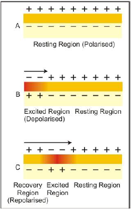
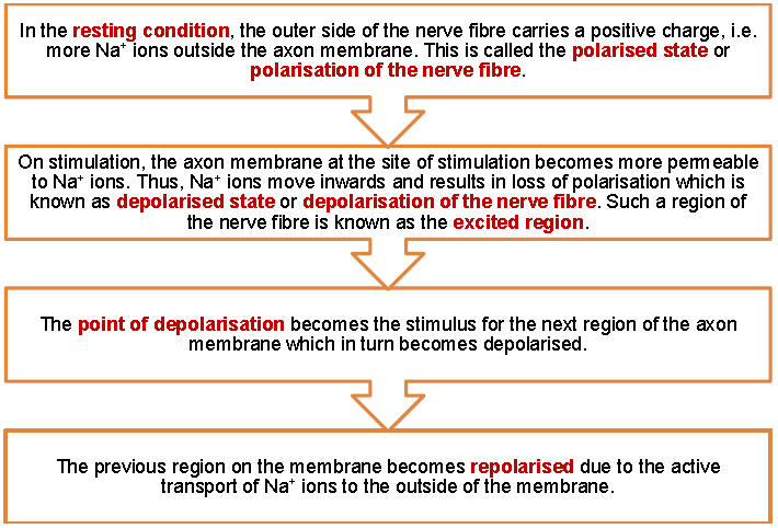
Under normal conditions, the outer side of the nerve fibre consists of positive charge as more Na+ ions are present outside axon membrane. The neuron is then said to be in polarised state. On stimulation, the membrane becomes more permeable and Na+ ions move inside causing depolarisation.
Such a region is known as excited region. The point of depolarisation behaves as stimulus for the neighbouring area and this goes on. In the mean time, the previous area becomes repolarised due to active transport (using ATP) of Na+ ions with the help of sodium pump.

Human Brain – Structure and Function
The body performs various activities. All these activities are controlled by the brain. How does the brain control all activities? Are there any divisions in the brain, which take over the control of different activities?
Do you know which organs make up the nervous system?
Let us explore.
The nervous system is divided into – central nervous system (CNS) and peripheral nervous system (PNS). The CNS consists of the brain and spinal chord while the PNS consists of the nerves that connect the central nervous system to different parts of the body.
The central nervous system receives information from all parts of the body and also sends information to the muscles. Communication between the CNS and body parts is facilitated by the nerves of PNS.
The important components of nervous system are:

The Central Nervous System
The central nervous system consists of the brain and the spinal cord. The brain is enclosed in a bony box called the cranium and spinal cord is protected by vertebral column.
The brain and spinal cord are externally covered by protective covering called meninges. It is made up of three layers namely duramater (outer layer), arachnoid (middle layer), piamater (inner layer).
The space between meninges is filled by a watery fluid called cerebro-spinal fluid (CSF). This fluid flows from the brain to spinal cord and then back to brain. It acts as a shock absorber and protects brain form injuries. It also provides nutrients to the cells in brain and spinal cord.
Human Brain
The brain is the main coordinating centre of the body. It is a part of the nervous system, which controls and monitors every organ of the body. The weight of the brain of an adult is about 1400 grams.
Different regions of the brain
The brain is divisible into three main regions—forebrain, midbrain, and hindbrain.

Forebrain
It is the main thinking part of the brain. It consists of the cerebrum, thalamus and hypothalamus. The forebrain has sensory regions, which receive sensory impulses from various receptors. It also has motor regions, which control the movement of various muscles such as leg muscles. There are separate areas in the forebrain specialized for hearing, smelling, seeing, general sensations such as pain, touch, taste, etc.
Cerebrum: The cerebrum is the largest part of the brain and constitutes four-fifth of its weight. It is divided by a deep cleft into two equal parts called left and right cerebral hemispheres.
Cerebrum has two regions, an outer cortex and inner medulla. The inner cortex is made up of cytons (nerve cell body) that give it a greyish appearance, so it is also called as grey matter. The medulla is composed of nerve fibres (axons and dendrites) that give it an opaque white appearance due to presence of myelin sheath covering, so is also called a white matter.
The cortex is provided with ridges called convolutions that increase the surface area of the cerebrum. The well developed cortex is responsible for the high degree of intelligence of the humans.
The information obtained through sense organs is stored in the cerebrum and used when needed. This ability to store information helps in retaining the memory.
A certain part of the cerebrum primarily controls intelligence, learning, memory, emotions, consciousness, thinking, and the ability to articulate speech. The forebrain is also known as the main thinking part of the brain.
In cerebrum, the nerves that come from the right side of the body are connected to the left side of cerebral hemisphere and the nerves that come from the left side of the body are connected to the right side of the cerebral hemisphere.
Therefore, organs of the right side of the body are controlled by left hemisphere and organs of the left side are controlled by the right hemisphere. Thus, injury in the left side of cerebral hemisphere results in the paralysis of organs on right side of the body and vice-versa.
Dienchephlon
It is the part of the forebrain located below the cerebrum. It includes both thalamus and hypothalamus.
Thalamus is situated between cerebral cortex and mid brain. It receives the nerve impulse form sense organs and transmits them to the upper region. It coordinates the sensory and motor signaling.
The hypothalamus contains many areas that control the body temperature, urge for eating and drinking, etc. Some regions of the cerebrum along with hypothalamus are involved in the regulation of sexual behaviour and expression of emotional reactions such as excitement, pleasure, fear, etc.
Midbrain
It is the small region of the brain that connects cerebrum with the hind brain. It has regions that are concerned with the sense of sight and hearing. Some regions of the midbrain transmit motor impulses to the limbs.
Hindbrain
It consists of three parts namely pons varoli, cerebellum and medulla oblongata.
Pons varoli consists of the nerve fibres that connect various portions like cerebrum, cerebellum and medulla oblongata of the brain. It has the control centers for facial expression, respiration and mastication etc. Among the twelve pairs of cranial nerves, four pairs originate from the pons varoli.
The cerebellum, which is a part of the hindbrain, is responsible for maintaining the posture and equilibrium of the body. It also coordinates the contraction of voluntary muscles, according to the directions of the cerebrum.
Medulla is the posterior most part of the brain and is connected to the spinal cord. Most involuntary actions such as heart beat, blood pressure, movement of food in the alimentary canal, salivation, etc. are controlled by the medulla of the hindbrain.
Spinal Cord
It is the continuation of the medulla oblongata and runs through the vertebral column. The spinal cord is made up of two similar halves fused together to form a central canal containing the cerebrospinal fluid. The outer portion of the spinal cord is known as the white matter, which consists of nerve fibres and the inner portion contains the cell bodies of neurons and is known as the grey matter.
There are thirty one pairs of spinal nerves that arise from the spinal cord. These nerves are divided into branches that reach to several parts of the body like, heart, lungs, stomach, urinary bladder, sex organs etc. The movement of limbs in the body are controlled by the spinal cord through reflex actions.
The spinal cord tapers at the end at the last vertebrae where from a collection of nerve roots originate, which are horsetail-like in appearance and hence called the cauda equina.

Protection to the brain and spinal cord
The brain, being an important organ, requires protection. Therefore, it is enclosed in a bony box called the cranium. The brain inside the brain box is also surrounded by a fluid-like material, which acts as shock absorber and thus, provides further protection to the brain. Spinal cord is protected by a bony, vertical rod with several curves called the vertebral column.


Peripheral Nervous System
It consists of the nerves arising from the brain and the spinal cord, which links the CNS to the rest of the body. It consists of two types of nerves.
• Cranial nerves: There are 12 pairs of cranial nerves and they emerge from the brain and reach the organs in the head region.
• Spinal nerves: There are 31 pairs of spinal nerves that emerge from the spinal cord and reach various parts of the body.
Messages are transferred from the brain to the spinal cord and then to the rest of the body and similarly messages from the rest of the body reach the spinal cord from where they are transferred to the brain. The spinal cord also controls all reflex actions.
Autonomic Nervous System
The autonomic nervous system helps to carry out the orders of the medulla, which controls the vital body functions.
It consists of two networks:
• Sympathetic system: The sympathetic nerves lead to all vital internal organs and glands. They regulate the actions of smooth muscles such as that of the stomach, intestine, and the heart.
• Parasympathetic system: This system is made up of the vagus and the pelvic nerves.
The sympathetic system speeds up the body functions and prepares the body for combat and escapes while the parasympathetic system counteracts to that of the sympathetic system and slows down the body functions.
Peripheral Nervous System and Autonomic Nervous System
Peripheral Nervous System (PNS)
PNS includes the nerves, which carry impulses to and from the CNS. Nerves included in the PNS are of two types- cranial nerves and spinal nerves.
Cranial Nerves
As the name suggests, these nerves originate from the cranium. Man has 12 pairs of cranial nerves, which include all three types of nerves − sensory, motor, and connector. The information about the 12 pairs of cranial nerves is given in the following table:


Spinal Nerves
• Spinal nerves are the nerves originating from the spinal cord by means of two roots- a dorsal root and a ventral root. • All the spinal nerves are connector nerves.
• At the junction of the two roots, the sensory nerve and motor nerves separate. The sensory nerves continue into the dorsal root and the motor fibres continue into the ventral root.
• Both the roots enter the grey matter of the spinal cord.
• Man has 31 pairs of spinal nerves, which are again put into five different categories:
• Cervical (8 pairs)
• Thoracic (12 pairs)
• Lumbar (5 pairs)
• Sacral (5 pairs)
• Coccygeal (1 pair)
Autonomic Nervous System
The nerves controlling the involuntary actions of the smooth muscles and glands when we are asleep or awake constitute the ANS.
ANS is present as chains of ganglion on either side of the backbone. Most of these ganglia are located close to or are embedded in the organ they control. Autonomic nervous system is divided into two systems − sympathetic and parasympathetic.
Sympathetic System − Sympathetic nerves originate from the thoraco-lumbar segment of the spinal cord. It gets activated during stressful conditions and stimulates the release of noradrenalin at the nerve endings. The main functions of the system are as follows:
• Dilation of iris
• Decrease in salivation (That is why our throat dries during stress)
• Increase in rate of heartbeat, dilation of bronchi
• Causes gylcogen breakdown to glucose in liver
• Inhibition of gastric and pancreatic activities
• Inhibition of peristalsis
Parasympathetic System − It gets activated during relaxation from stress. All the effects of its activation are opposite to sympathetic system. Therefore, we can say that these two systems are antagonistic to each other.
Responses of the Nervous System
What happens when the following takes place?
• Bright light is focused on our eyes
• We accidentally touch a flame
• We are hungry and we think about our favourite meal For
all the situations mentioned above, the response would be quick and automatic. We would
• close our eyes immediately when bright light is focused on our eyes
• withdraw our hand from the flame
• start salivating on thinking about our favourite meal
This automatic action or response provoked by a stimulus is known as a reflex action.
The responses of the nervous system can be classified into voluntary, involuntary, and reflex actions.
The actions that can be controlled voluntarily are called voluntary actions. The signal or message for these actions is passed to the brain. Therefore, they are consciously controlled.
On the other hand, the movement of food in the alimentary canal or the contraction and relaxation of the blood vessels are involuntary actions i.e.they cannot be consciously controlled.
The reflex actions, however, show sudden responses and do not involve any thinking. This means that unlike involuntary actions, these actions are not under the control of the brain.
Reflex arc
When we accidentally touch a hot object, we withdraw our hands immediately without thinking. If we do not do this, our hands will burn.
The sensory nerves detect the heat. They are connected to the nerves, which move the muscles of the hand. Such a connection of detecting the signal from the nerves (input), and responding to it immediately (output) is called a reflex arc. In other words it is the pathway along which nerve impulse travels during the reflex action.
A reflex arc makes instant and automatic responses possible. It connects the input nerve and output nerve, and meets in a bundle in the spinal chord. In fact, nerves from all over the body meet in a bundle in the spinal cord, on their way to the brain. Therefore, the information input reaches the brain.

The reflex arc consists of five distinct parts and these are:
1. Receptor: It includes sense organs that receive stimulus.
2. Sensory neuron: It conducts the nerve impulse from receptor to the spinal cord or brain.
3. Association neuron: It helps to transmit nerve impulse from sensory neuron to motor neuron.
4. Motor neuron: It transmits nerve impulse to the effector organs like muscles or glands.
5. Effector: It includes muscles or glands where action takes place in response to stimulus.
Types of Reflexes
Ivan Pavlov classified all reflex responses in two categories − Unconditional and conditional reflexes.
Unconditional Reflexes − These are the inborn, unconscious responses to a given stimuli which are transferred to the next generation as well.
Some of the examples of such unconditional responses are suckling of the mother’s breast by a new born body blinking of eyes when an object is brought very close to the eyes.
Condition Reflexes − Such responses are acquired during the life time of an individual. These responses are different for different organisms. These responses can be easily induced or lost depending upon the environmental conditions.
Pavlov’s Experiment on a Dog In this experiment the Russian famous biologist, Ivan Pavlov tested the conditional reflexes. He used a dog as his experiment subject and tested for the secretion of saliva in response to ringing of a bell.
Under normal condition, dog will not secret saliva on listening the ringing of a bell or any other sound. In his experiment, Pavlov brought food and rang the bell simultaneously for a prolonged period of time.
After an adequate period of training, it was observed that the dog started secreting saliva just by listening to the bell’s ringing.

Conditional reflexes are controlled by cerebral cortex.
Some of the examples of conditional or acquired reflex are learning, playing piano, typing on a computer, etc.
Structure of Human Eye
Sense Organs: Organs that helps us to be aware of our surroundings are known as sense organs. Some of the major sense organs of our body include eyes, ears, nose, tongue and skin.
Receptors: Any cell or tissue sensitive to a selective stimuli is known as receptor. Some common receptors are:
• Mechanoreceptors: Receptors for touch or pressure; found in skin
• Thermoreceptors: Receptors fro temperature; found in skin
• Chemoreceptors: Receptors of taste (in tongue) and of smell (in nose)
• Photoreceptors: Receptors of light; found in eyes (rod and cone cells)
Eye
Eye is one of the most sensitive sense organs in the human body. Our eye enables us to see this beautiful world. It consists of a lens, which is made up of living tissues. How does our eye work? What are the nature, position and relative sizes of the images formed by the lens in the eye? In this section, we will learn about the structure and functioning of human eye.
Structure of human eye

The human eye is roughly spherical in shape with diameter of about 2.3 cm. It is situated in the front side of the skull in bony sockets. It is covered by the eyelids that have eye lashes which prevent dust and other substances from entering into the eye. It consists of a convex lens made up of living tissues.
Hence, human lenses are living organs contrary to the simple optical lenses. The inner region of the upper eye lid contains Lacrymal glands, that produces secretion known as tears, which keep eye surface moist and wash out dirt and other substances. Tears contain some salts and act as an antiseptic because of the presence of the enzyme lysozyme which kills the germs.
Structure of Human Eye:
The wall of the eye consists of three layers namely sclera, choroid and retina.
The following table lists the main parts of the human eye and their respective functions.
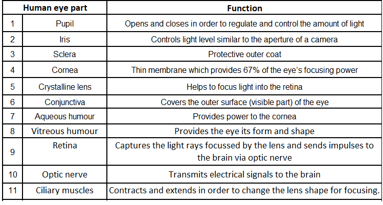
The white of the eye is known as the sclera. It is the tough, opaque tissue that protects the outer layer of the eye. The bulged, transparent front portion of the sclera is called cornea. It is protected by thin, transparent tissue known as the conjunctiva.
The middle layer called choroid is supplied with nerves and blood vessels. It consists of the coloured layer of tissue called iris. It is responsible for the colour of the eye. Pupil is the black, circular hole that is located at the centre of the iris.

The lens consists of layers of tissues enclosed in a tough capsule. The focus of the lens is adjusted by the ciliary muscles that suspend and hold it. The lens focuses the light rays on retina where inverted image of the object is formed.
Retina contains two types of cells rods and cones. Rods are sensitive to dim light and cannot differentiate between various colours while as cones are sensitive to bright light and can distinguish various colours.
Functioning of the human eye
Light rays enter the eye through the cornea. The rays are bent, refracted, and focused by the cornea, lens, and the vitreous humour. The main function of the lens is to focus
the light rays sharply on the retina. It is the outer surface of the cornea where most of the refraction of light occurs.
Iris controls the size of the pupil and the amount of light respectively. Since the eye lens is convex in nature, the resulting image is real, small, and inverted. This image is formed on the retina.
The retina converts these light rays into electrical signals with the help of light sensitive cells. These signals are sent to the brain via translated and perceived objects in an erect or upright position.
The head of the optic nerve is devoid of photosensitive cells (rods and cones). Hence, no image is formed at that point called the blind spot of the eye.
Lateral to the blind spot, a yellow spot (fovea) is present that contains large number of cone cells. At this portion of retina a most clear and sharp image is formed.
Power of Accommodation
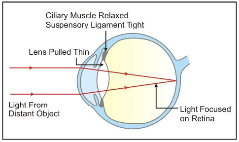
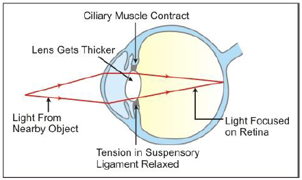
This is a special capacity of human eye to adjust its focus depending upon the object they are seeing. This happens because of the presence of flexible ciliary muscles around the eyes that helps in adjusting the focus of the eye lens. For distant vision the lens flattens whereas for near vision it becomes more convex.
Stereoscopic Vision
Humans and monkeys/apes have a special ability to perceive depth and relative distance as they can simultaneously focus on an object with both eyes. This results in the generation of a three dimensional image in our brain. This ability is known as stereoscopic vision.
On sunny days, when you enter a dimly lit room, you are unable to see clearly for a moment. Why does this happen?
In bright light, the iris expands, thereby contracting the pupil. This happens so that only a small quantity of light enters the eye. As a result, the retina is protected from exposure to excessive light.
On entering a dimly lit room after having been in the sun for some time, the iris contracts slowly to expand the pupil. Gradually, more light is able to enter the eye. Hence, it takes a few seconds before we are able to see the objects present in the dimly lit room.
Common Defects of Eye


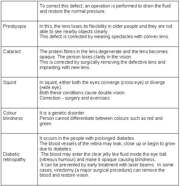
Let’s study a defect of eye known as presbyopia through this animation.
First Aid for the Removal of Foreign Bodies from the Eyes
• In case the foreign bodies like dust particles, flies, metal particles or saw dust of wood etc gets into the eyes, do not rub the eyes as it may cause injury to the eye ball.
• If foreign body is visisble, remove it with the help of clean and soft cloth.
• It can be also removed by pulling one eye lid over the other that results in the flow of tears. The flow of tears help to wipe the foreign particles out.
• It can be also removed by keeping the eyes in bowl of cold water or boric lotion and closing and opening the eyes several times. This also helps to wipe out the foreign paticles.
Structure of Human
Ear Functions of the Ears
• Hearing
• Maintenance of body balance
Anatomy of the Ear
• Divided into three major sections:
• Outer ear
• Middle ear
• Inner ear

• Outer ear = Pinna + External auditory meatus (Canal)
• Pinna − collects the vibrations that produce sound
• Canal − leads inwards and extends up to the tympanic membrane (Ear drum)
• Wax-secreting sebaceous glands are present in the skin of the pinna and the canal.
• Middle ear: Has 3 ossicles (Malleus, Incus, Stapes)
• Malleus is attached to the ear drum and stapes is attached to the oval window of the cochlea. Middle ear communicates to the inner ear through the oval window
• Ossicles increase efficiency of transmission of sound waves to the inner ear.
• Eustachian tube − connects the middle ear cavity with the pharynx; equalises the pressure on either sides of the ear drum
• Inner ear (labyrinth): Has 2 parts (Bony Labyrinth and Membranous Labyrinth)
• Bony Labyrinth − series of channels in which the membranous labyrinth lies
• Membranous Labyrinth − surrounded by a fluid called perilymph and filled with a fluid called endolymph
• Cochlea − coiled portion of the labyrinth
• 2 membranes surround cochlea, the reissner’s membrane and the basilar membrane.
• These membranes divide the bony labyrinth into 3 parts -upper scala vestibuli, middle scala media and lower scala tympani.
• Scala media − filled with endolymph
• Scala vestibuli − ends at the oval window Scala tympani − ends at the round window that opens to the middle ear
• Organ of Corti − located on the basilar membrane and contains auditory receptors called hair cells (close contact with afferent nerve fibres)
• Tectorial membrane − elastic membrane present above the rows of hair cells

• Vestibular apparatus: Complex system located above the cochlea
• Composition − 3 semi-circular canals and otolith organ (has saccule and utricle)
• Base of the canals is swollen to form ampulla, which contains a projecting ridge called the crista ampullaris; it has hair cells
• Saccule and utricle − contain macula, which along with crista is responsible for maintaining the balance of the body and posture
Mechanism of Hearing
Role of Ear in Body Balancing
The fluid inside the semicircular canals moves when we turn our head. This moving fluid pushes against the sensory hair cells. This results in transmission of nerve impulse to brain through auditory nerve. The cells present in the semicircular canals are highly sensitive to dynamic equilibrium. Hence, they help us in maintaining balance of our bodies.
Senses of Smell, Touch and Taste
Sense Organs, in humans and other animals, are the faculties by which outside information is received for evaluation and response. This is accomplished by the effect of a particular stimulus on a specialized organ, which then transmits impulses to the brain via a nerve or nerves.
There are five type of senses which can be sensed by our sense organs – touch, vision, hearing, smell and taste.
These five type of senses can be categorised as general and special senses.
• General senses –
• contain general sensory receptors
• mostly modified dendritic ends of sensory neurons
• present throughout the body
• monitor most of the types of general sensory information such as tactile sensation, heat, cold, pain and muscle sense • Special senses –
• contain special sensory receptors
• confined to the head region, sensory organs like eyes and ears and tissues of the taste buds and olfactory epithelium We are very much familiar with eye and ear. So let’s learn the details of other three sense organs.
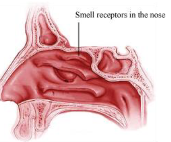
Nose
The nose is the organ for smelling. The receptors for smell called olfactory receptors are present in the nose to perceive the smell. Impulses are sent to the brain by these receptors and we are able to smell things
• The receptors for smell are located in small patches is the upper portion of the nasal cavity. The mucosa of upper nasal chamber is called olfactory epithelium.
• Three types of cells present in olfactory epithelium- olfactory receptor cells, columnar supportive cells and short basal cells.
• Olfactory receptors are unusual bipolar sensory neurons.
• Thin dendrites of neurons bear clusters of 20 modified cilia which function as receptor sites.
• Cilia extend from the olfactory epithelium into the thin coat of nasal mucous secreted by the supportive cells and olfactory glands.
• Mucus present in nose acts as a solvent.
• The olfactory nerve carries the stimuli from these cells to the brain.
• The smell receptors in the nose are sensitive to chemicals and they send impulses to the brain via the olfactory nerve to the cerebrum.
Tongue
The tongue is the sense organ responsible for taste. It has patches of sensory receptors known as taste buds. Different portions of the tongue are responsible for comprehending the different tastes.
• Nerves arise from the ends of the sensory taste buds and they together constitute the taste nerve.
• The nerve carries the signals to the brain where it is interpreted.
• The tongue has taste buds for the four basic tastes − sweet, sour, bitter and salty.
• The tip of the tongue is sensitive to sweet and salty tastes.
• The sides are sensitive to sour taste.
• The posterior portion is sensitive to bitter taste.

• Mechanism of tasting –
• dissolved chemicals contacting the microvilli bind to specific receptor proteins on it
• this depolarizes the cell
• dendrites of the associated sensory neurons coil intimately around the receptor cells and synapse with them
• a neurotransmitter is released when a receptor cell is stimulated and depolarized
• this leads to the generation of an action potential in the associated sensory neuron
• each dendrite receives signals from several receptor cells within the taste bud
• nerve fibers emerging from the taste buds pass to the brain stem
• from here the nerve impulse is relayed to the taste centre in the cerebral cortex of the brain that perceives the taste sensation
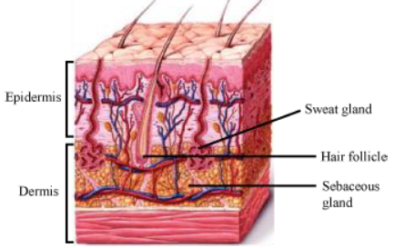
Skin
The skin is the sense organ for touch and feel. It also protects the body. It makes us aware of temperature, pressure, pain, etc
• Skin has two layers –
• The epidermis − It is the outer layer, made up of epithelial tissue and contains a brown pigment called melanin.
• Dermis − It is the inner layer and is made up of connective tissue and contains sweat glands and oil glands which give off moisture and oil which keeps the skin soft.
• It also contains hair follicles from which hair grows the skin also has nerve endings which act as touch receptors.
• Some of the receptors present in the skin are

Lets study some of these receptors in detail –
• Free or bare dendritic nerve endings – present throughout the epidermis taking an extensive branching or “zigzag” form. Respond chiefly to pain and temperature but some respond to pressure as well.
• Meissner’s corpuscles – small receptors in which a few spiraling dendrites are surrounded by specialized capsule (Schawann) cells. Found just beneath the skin epidermis in dermal papillae and especially abundant in finger tips and soles of the feet.
• Pacinian corpuscles – large egg shaped bodies. Single dendrite surrounded by multilayers of capsule cells. Scattered deep in the dermis and in the subcutaneous tissue of the skin.
Mechanism of touch –
• an impulse or action potential is generated whenever one or more of these sensory receptors are stimulated (by heat, cold, vibrations, pressure or pain)
• impulse is then taken to the spinal cord and from there to the brain
• brain analyses the stimulus and then generates an appropriate response
Our nervous system consists of the brain, spinal cord, sense receptors and nerves.
Functions of the Nervous System
• Keeps us informed about the outside world through sensory organs.
• Controls and harmonises all voluntary muscular activities, e.g. running and writing.
• Enables us to remember, think and reason.
Neuron: The Unit of the Nervous System
Structure of the Neuron

The three main parts of the neuron are as follows:
• Cell Body: It has a well-defined nucleus and granular cytoplasm.
• Dendrites: They are the branched cytoplasmic projections of the cell body.
• Axon:
o It is a long process of the cell body.
o The axon is covered by a myelin sheath.
o The myelin sheath shows gaps throughout its length known as Nodes of Ranvier.
Some Basic Terms
Stimulus: An agent or sudden changes of the external or internal environment which results in a change in an organism or any of its body parts. Response: The change in organisms resulting from a stimulus.
Impulse: A wave of irritability, i.e. an electrical disturbance, which sweeps over the nerve cell. Receptors: The nerve cells which set up waves of impulses towards the central nervous system on receiving the stimulus.
Effectors: Muscles or glands which contract or secrete substances on receiving an impulse from the brain or the spinal cord
Synapse
• A synapse is the point of contact between the terminal branches of the axon of a neuron and the dendrites of another neuron.
• As the nerve impulse reaches the axon terminal of one neuron, the neurotransmitter acetylcholine is released by the bulbs present in the axon.
• Acetylcholine is then broken down by an enzyme to ensure that the synapse is ready for the transmission of the next nerve impulse.

Transmission of Nerve Impulse
Types of Neurons
• Sensory Neurons: Convey the impulse from the receptors (sense organs) to the main nervous system (the brain or spinal cord).
• Motor Neurons: Carry impulse from the main nervous system to an effector, i.e. muscle or gland.
• Associated Neurons: They interconnect sensory and motor neurons.
Types of Nerves
A nerve is a bundle of nerve fibres (axons) of separate neurons enclosed in a tubular sheath. Ganglia are an aggregation of the nerve cells (cell bodies) from which the nerve fibres may arise or enter.
Division of the Nervous System
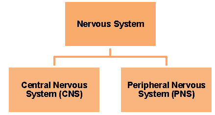
The Central Nervous System
The central nervous system includes the brain and the spinal cord.
The Brain
• The human brain is well protected inside the cranium or the skull.
• In adults, it weighs about 1.35 kg
• It is protected by three meninges—dura mater, arachnoid and pia mater.
• The space between the covering membranes, central spaces of the brain and the central canal of the spinal cord consists of cerebrospinal fluid which protects the brain from shocks.
Three Primary Regions of the Brain
• Forebrain
o The cerebrum is the centre of intelligence, memory, consciousness, will power and voluntary actions.
o The thalamus relays pain and pressure impulses to the cerebrum.
o The hypothalamus controls the body temperature and the activity of the pituitary gland.
• Midbrain
o This small tube-like part is responsible for reflexes involving the eyes and ears.
• Hind Brain
o The cerebellum coordinates muscular activity and balance of the body.
o The pons carries impulses from one hemisphere to the other hemisphere and coordinates muscular movements on both sides of the body.
o The medulla oblongata controls the activities of internal organs, heartbeat, breathing etc.
Parts of the Brain


The Spinal Cord

• Lies within the neural canal of the vertebrae.
• The grey matter is on the inner side and the white matter is on the outer side of the spinal cord.
• Similar to the brain, it is covered with three meninges—dura mater, arachnoid and pia mater.
• Functions:
o Responsible for reflexes below the neck.
o Conducts sensory impulses from the skin and muscles to the brain.
o Conducts motor responses from the brain to muscles of the trunk and limbs.
Peripheral Nervous System
The peripheral nervous system consists of nerves which carry impulses to and from the central nervous system.

Somatic Nervous System
• Cranial Nerves: 12 pairs emerge from the brain.
• Spinal Nerves: 31 pairs: 8 pairs in the neck region, 12 pairs in the thorax, 5 pairs in the lumbar region, 5 pairs in the sacral region and 1 pair in the coccygeal region.
Autonomic Nervous System
The autonomic nervous system controls the involuntary actions of the internal organs.

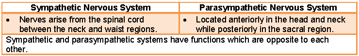
Opposite Effects of the Two Systems

Reflexes
The reflex action is an automatic, quick and involuntary action in the body brought about by a stimulus. Difference between Reflexes/Involuntary Actions and Voluntary Actions

Some examples of reflexes:
• Shivering when it is too cold or sweating when it is too hot.
• Non-stop beating of the heart.
• Instant withdrawal of the hand when it accidently touches a hot iron.
• Dilation of the pupil in eyes when looking in the dark.
Types of Reflexes
Pavlov’s Experiment

Nervous Pathways in Reflexes A reflex
action must be quick to give quick response. Therefore, the pathway for receiving and sending information must be short. A reflex arc can be represented as follows: Stimulus → receptor in the sense organs → afferent (sensory) nerve fibre → CNS (spinal cord/brain) → efferent (motor) nerve fibre → muscle/gland → Response
The Sense Organs
The sense organs enable us to be aware of the condition of the environment. A receptor is any specialised tissue or cell sensitive to a specific stimulus.

The Eyes
• The two eyes are located in deep sockets called orbits.
• The upper and lower moveable eyelids protect the front surface of the eyes.
• There are 6–12 tear glands.
• Functions of the tear glands are
o Lubricate the surface of the eye
o Wash away the dust particles
• A thin membrane which covers the entire front part of the eyes is called conjunctiva.
• Due to viral infection of the conjunctiva, we suffer from eye disease called conjunctivitis.
Structure of the Eyeball
The wall of the eyeball is composed of the following three concentric layers:
1. Sclerotic Layer (Outer Layer)
o The white visible portion of the eyeball is nothing but the sclera.
o The sclera covers the coloured part of the eye, i.e. the cornea.
2. Choroid Layer (Middle Layer)
o Richly supplied with blood vessels to provide proper nourishment.
o Choroid expands in the front to form a ciliary body.
o Iris is also a part of the choroid.
o The iris partially covers the lens. It leaves a circular opening in the centre called a pupil.
o The muscles of the iris regulate the size of the pupil. Thus, the pupil regulates the amount of light entering the eye.
3. Retina (Inner Layer)
o It has two types of sense cells—rods and cones.
o The rod cells are sensitive to dim light and do not respond to colour.
o The cone cells are sensitive to bright light and are responsible for colour vision.
Comparison between Rods and Cones
Yellow Spot and Blind Spot
Lens
• It is transparent, biconvex and crystalline.
• It is held by a suspensory ligament which attaches the lens to the ciliary body.
Aqueous and Vitreous Chambers
The lens divides the inner cavity of the eye ball into two chambers:
Four Major Steps in Seeing an Object
Accommodation Vision
The process of focusing the eyes at different distances is called accommodation. This is brought about by change in the curvature of the lens.
• For distant vision, the lens is more flattened.
The lens remains stretched by the suspensory ligaments.
• For near vision, the lens becomes convex and rounded.
The ciliary muscles contract and pull the ciliary body forward. This releases the tension of suspensory ligaments, making the lens convex and rounded.
Light and Dark Adaptation
Dark Adaptation
When we pass from a brightly lit area to a dark area, we experience difficulty in seeing the objects for a short while. This is called dark adaptation
Light Adaptation
When we pass from a dark area to a brightly lit area, we experience a dazzling effect for a short period. This is called light adaptation.
Common Defects of the Eyes

Stereoscopic Vision
Humans, monkeys and apes can perceive depth or the relative distance of objects. This is due to simultaneous focusing of an object in both eyes. The images of both eyes are overlapping and give a 3-dimensional effect.
After-images
When one looks at a brightly coloured object and then looks at a dark surface, an image of the object in the same colour will persist. This is known as persistence image or after-image.
The Ear
The human ear has the three following main divisions:

1. Outer Ear
• Consists of pinna/auricle and auditory canal.
2. Middle Ear
• Contains three ear ossicles—malleus (hammer), incus (anvil) and stapes (stirrup)—and the eustachian tube.
• The eustachian tube connects the cavity of the middle ear with the throat.
3. Inner Ear
• Also known as membranous labyrinth.
• Contains cochlea and the semicircular canals.
• The cavity of cochlea is divided into three parallel canals. The middle canal consists of the organ of corti which is responsible for hearing.
• Ends of the semicircular canals widen to form an ampulla.
• The ampulla contains sensory cells.
• The short stem joining the bases of semicircular canals to the cochlea is called the vestibule.
• The vestibule contains two sacs—utriculus and sacculus.
Functions of the Ear
Two functions of the ears are hearing and body balance.
1. Hearing
The pinna collects sound waves and conducts them through the external auditory canal. They finally strike on the ear drum and the vibration is set.

2. Body Balance
• The sensory cells in the semicircular canals are concerned with dynamic equilibrium, i.e. when the body is in motion.
• The sensory cells in utriculus and sacculus are concerned with static equilibrium, i.e. when the body is stationary.
Hearing Impairment

The Sense of Taste

• The sense of taste is located in the taste buds of the tongue.
• A taste bud is an ovoid group of sensory cells.
• Substances enter the pore and stimulate the sensory hair of the sensory cells.
The Sense of Smell
• The sense of smell is located in the epithelial layer of the nasal chamber.
• The sense cells for smell have hair-like projections.
• These hair-like projections respond to particles dissolved in the mucous secretion of the nose.
• The impulse from these cells is then transmitted to the brain via the olfactory nerve.