Multiple Choice Type Questions:
(Select the most appropriate option in each case)
Question 1: Agranulocytes are:
(a) lymphocytes and monocytes
(b) lymphocytes and basophils
(c) eosinophils and basophils
(d) eosinophils and monocytes
Solution :
lymphocytes and monocytes
Question 2: White blood cells engulf bacteria in a process called:
(a) diapedesis
(b) phagocytosis
(c) active transport
(d) passive transport
Solution :
phagocytosis
Very Short Answer Type Questions:
Question 1: Given below are certain structures, write the term for the functional activity.
(a) Blood platelets and …………………………….
(b) Neutrophils and …………………………………..
(c) Erythrocytes and …………………………………
(d) Lymphocytes and …………………………………
(e) Bone marrow and ………………………………..
Solution 1:
(a) Blood platelets and blood coagulation
(b) Neutrophils and phagocytosis
(c) Erythrocytes and transportation of gases
(d) Lymphocytes and Produce antibodies
(e) Bone marrow and destruction of old and weak RBC’s/production of RBCs and WBCs.
Question 2: Name the following:
(a) The cells which transport oxygen to the different parts of the human body.
(b) The cells that initiate blood clotting.
Solution 2:
(a) Red Blood Cells
(b) Blood Platelets
Short Answer Type Questions:
Question 1: Enumerate the structural differences between white blood cells and red blood cells.
Solution 1:
Structural Differences between White Blood Cells and Red Blood Cells:
| White Blood Cells | Red Blood Cells |
| White blood cells are amoeboid in nature. | Red blood cells are biconcave disc-like structures with a biconcave shape. |
| They are nucleated cells, which means they have a nucleus. | Cells were anucleated. |
| White blood cells do not contain haemoglobin. | Red blood cells contain haemoglobin. |
Question 2: Why is it necessary to know the blood groups before giving transfusion?
Solution 2:
Before a blood transfusion, it is crucial to know the blood groups of both the donor and the recipient since the blood groups of the donor and recipient must be compatible. In the instance of an incompatible blood transfusion, the recipient produces antibodies that attack the antigens on the donor’s RBCs, causing the blood cells to clump together and potentially cause death.
Question 3: Differentiate between members of each of the following pairs with reference to phrases in brackets:
(a) Antibodies and antibiotics (Source)
(b) RBC and WBC (structure)
(c) serum and vaccine (Composition)
Solution 3:
(a) Antibodies: Lymphocytes generate antibodies in response to infections entering the bloodstream.
Antibiotics: Antibiotics are antibiotics derived from bacteria and fungi. Antibiotics kill or stop infections from growing.
(b) RBC: RBC is an enucleated, biconcave, disc-like structure with a flat centre and thick, rounded perimeter. WBC: WBC is a nucleated, amoeboid-shaped blood cell.
(c) Serum: Serum is plasma that has had the protein fibrinogen removed from it.
Vaccine: Vaccines are dead or weakened microorganisms that are delivered into the body in order to encourage the creation of antibodies against pathogens that cause a certain disease.
Question 4: Complete the following statement by filling in the blank from the choices given in the brackets. An anticoagulant present in the blood is (heparin, hirudin, thromboplastin, calcium).
Solution 4:
Heparin
Long Answer Type Questions:
Question 1: What are the functions of blood plasma?
Solution 1:
The functions of blood plasma are:
(a) Transports of digested food from the gastrointestinal tract to the tissues.
(b) Moves excretory materials from the tissues to the excretory organs.
(c) Transports hormones from the glands to their destination.
(d) Maintains body temperature by distributing heat throughout the body.
Question 2: What are the main steps in coagulation of blood?
Solution 2:
The following steps are involved in blood clotting or coagulation:
a) At the wound site, wounded tissue cells and platelets dissolve, releasing thromboplastin.
b) Thromboplastin turns inactive prothrombin into active thrombin with the help of calcium ions.
c) Thrombin transforms soluble fibrinogen into insoluble fibrin in the presence of calcium ions, forming a mesh or network at the wound site.
d) The blood cells stuck in this network contract and squeeze out the plasma, leaving a solid mass known as a clot behind.

Question 3: What are the following?
a) Rh factor
b) Universal donor
c) Diapedesis.
Solution 3:
(a) Rh factor – Rh factor is a hereditary antigen that is frequently seen on blood cells. Some people have these antigens and are thus Rh positive (Rh+), whereas others do not and are thus Rh negative (Rh-) (Rh-).
(b) Universal donor – A person with blood group O is a universal donor since this blood type can be given to people with any blood group, including O, A, B, and AB.
(c) Diapedesis – Diapedesis is the squeeze of leucocytes through capillary walls into tissues.
Question 4: Is it possible for the blood to clot under the skin? Give reason in support of your answer.
Solution 4:
The exposure of blood to air has no effect on blood coagulation. The passage of blood over a rough surface, such as a cholesterol deposit inside a blood vessel of the skin, can actually cause clotting.
Question 5: State any five functions of the blood.
Solution 5:
The blood has the following functions:
(a) Transporting digested food from the alimentary canal to the tissues. Simple sugars like glucose, amino acids, vitamins, mineral salts, and other compounds fall into this category.
(b) Transport of oxygen from the lungs to the tissues in the form of the unstable molecule oxyhaemoglobin.
(c) Carbon dioxide transport from the tissues to the lungs.
(d) Excretory contents are transported from the tissues to the liver, kidney, or skin for evacuation.
(e) Hormone distribution from glands to target areas.
(f) Heat distribution to maintain a constant body temperature.
Structured / Application / Skill Type Questions:
Question 1: Given below is a diagram of a smear of human blood. Study the same and answer the questions that follow:
(a) Name the parts 1, 2, 3 and 4 indicated by guidelines.
(b) Mention two structural differences between the parts labelled ‘1’ and ‘2’.
(c) What is the main function of the parts labelled 1, 2, 3 respectively?
(d) What is the life span of the part labelled ‘1’?
(e) Name a soluble protein found in ‘4’ which helps in the clotting of blood.
Solution 1:
(a)
i) Red Blood Cell (RBC),
ii) White Blood Cell (WBC),
iii) Blood Platelet
iv) Blood Plasma.
(b) Red blood cells resemble little biconcave discs, whereas white blood cells are amoeboid.
(c) Function of portion 1 (RBC): Transport of breathing gases to and from tissues, as well as nutrients from the alimentary canal to tissues.
Function of part 2 (WBC): WBCs serve a critical function in the body’s defence and immunity mechanisms.
Part 3 (Blood Platelet) has the following function: Blood platelets are the catalysts for blood coagulation.
(d) A red blood cell (RBC) has a lifespan of roughly 120 days.
(e) Thromboplastin
Multiple Choice Type:
(Select The Most Appropriate Option In Each Case)
Question 1: The nearest organ to which the heart supplies oxygenated blood is:
(a) Lung
(b) Stomach
(c) Intestine
(d) Heart itself
Solution :
Heart itself
Question 2: When a doctor is recording your pulse, he is pressing on your wrist exactly on a
(a) vein
(b) capillary
(c) artery
(d) arteriole
Solution :
artery
Question 3: The valve present between the right atrium and the right ventricle is the
(a) tricuspid valve
(b) bicuspid valve
(c) semi-lunar valve
(d) mitral valve
Solution :
tricuspid valve
Question 4: The blood vessel supplying blood to the kidney is the
(a) renal vein
(b) renal artery
(c) dorsal aorta
(d) hepatic vein
Solution :
renal artery
Question 5: Angina pectoris is due to
(a) defective nutrition
(b) inadequate supply of oxygen to the heart muscle
(c) defective functioning of mitral
(d) infection by a virus
Solution :
inadequate supply of oxygen to the heart muscle
Question 6: The chief function of lymph nodes is to
(a) produce WBCs
(b) produce hormones
(c) destroy old RBCs
(d) destroy pathogens
Solution :
destroy pathogens
Question 7: Heart sounds are produced due to
(a) Closure of tricuspid and bicuspid valves
(b) Rushing of blood through valves producing turbulence
(c) Closure of aortic and pulmonary valves
(d) Entry of blood into auricles
Solution 7:
(a) closure of tricuspid and bicuspid valves
(b) closure of aortic and pulmonary valves
(c) rushing of blood through valves producing turbulence
Very Short Answer Type Questions:
Question 1: What are the average values of blood pressure in a normal adult human?
Solution 1:
Blood pressure in a healthy adult person ranges from 100 to 140 mm for systolic pressure and 60 to 80 mm for diastolic pressure.
Question 2: Is it true that your heart beats more than one lac times per day?
Solution 2:
Yes, the heart beats around 1,036,680 times every day.
Question 3: Name the following:
a) Any one vein which starts from an organ and ends in another organ besides the heart.
b) The kind of blood vessels which have no muscular walls.
c) An artery which carries impure (deoxygenated) blood.
d) The kind of blood cells which can squeeze out through the walls of one category of blood vessels.
e) The smallest common blood vessels formed by the union of capillaries.
f) The blood vessels which start from capillaries and end in capillaries.
g) The phase of the cardiac cycle in which the auricles contract.
h) The valve present in between the chambers on the right side of the human heart.
i) The phase of the cardiac cycle in which the ventricles get filled with blood from the atrium.
j) The fluid found between the membranes of the heart.
Solution 3:
a) Hepatic portal vein
b) Blood Capillaries
c) Pulmonary artery
d) White blood cells
e) Venules
f) Portal vein
g) Atrial systole
h) Tricuspid valve
i) Atrial systole
j) Pericardial fluid
Question 4: Write the chief functional activity of each of the following:
(a) Blood platelets …………………………..
(b) Neutrophils ………………………………
(c) Erythrocytes …………………………….
(d) Lymphocytes ……………………………
(e) Bone marrow ……………………………
Solution 4:
(a) Blood platelets have a role in clotting and coagulation. At the site of injury, blood platelets combine to release thromboplastin, which starts the clotting process.
(b) Neutrophils engage in phagocytosis, which means they engulf and eliminate pathogens that enter the bloodstream.
(c) Erythrocytes transport oxygen in the form of the unstable molecule oxyhaemoglobin from the lungs to the tissues and carbon dioxide from the tissues to the lungs.
(d) Antibodies are produced by lymphocytes in response to infections that enter the bloodstream. They may also do phagocytosis in some situations.
(e) The development of RBCS and WBCs is aided by bone marrow. It’s also responsible for destroying aged and weak RBCs.
Question 5: Complete the following statements by filling in the blanks from the choices given in the brackets.
(a) The blood vessels that begins and ends in capillaries is the (hepatic artery,
hepatic portal vein, hepatic vein)
(b) A blood vessels which has small lumen and thick wall is (capillary,
lymphatic duct, artery, venule)
(c) The valve which prevents back flow of blood in the veins and lymph vessels ………………
(mitral valve, tricuspid valve, semilunar valve).
Solution 5:
(a) The blood vessel that begins and ends in capillaries is the hepatic portal vein.
(b) A blood vessel which has small lumen and thick wall is artery.
(c) The valve which prevents the back flow of blood in the veins and lymph vessels is semilunar valve.
Question 6: Note the relationship between the first two words and suggest the suitable word/ words for the fourth place:
(a) Lubb: Atrioventricualr valves :: dup: …………………
(b) Coronary artery: Heart :: Hepatic artery : ……………
Solution 6:
(a) Lubb : Atrio-ventricular valve :: Dup : Semilunar valves
(b) Coronary artery : Heart :: Hepatic artery : Liver
Question 7: Give reason, why a matured mammalian erythrocyte lacks nucleus and mitochondria?
Solution 7:
The nucleus and mitochondria of a mature mammalian erythrocyte are missing. RBCs with no nucleus have a higher surface area-to-volume ratio, which improves the surface area available for oxygen absorption. Furthermore, the absence of a nucleus lowers the size of the cell, making it easier to flow through blood channels and allowing more cells to fit into a compact space.
Because the cell lacks mitochondria, it does not consume any of the oxygen taken for respiration, enhancing the cell’s oxygen transport efficiency because all of the oxygen absorbed is transferred without loss.
Short Answer Type Questions:
Question 1: What does the term “double circulation” mean?
Solution 1:
Before it completes one full round, blood passes twice through the heart. Pulmonary and systemic circulation are thus included in the whole circle. Blood flows into the lungs via pulmonary arteries in pulmonary circulation. The blood from the lungs is collected and carried back to the left atrium by pulmonary veins.
Blood from the left ventricle enters the aorta, which then transports the blood to the other bodily sections. Veins gather blood from various bodily parts and return it to the heart. As a result, the human body’s blood circulation is referred to as twofold circulation.
Question 2: When are the sounds “LUBB” and “DUP” produced respectively during heart beat?
Solution 2:
When the atrio-ventricular valves, namely the tricuspid and bicuspid valves, close at the onset of ventricular systole, the first sound, LUBB, is created.
When the pulmonary and aortic semilunar valves close at the start of ventricular diastole, the second sound DUP is created.
Question 3: Why do people have a common belief that the heart is located on the left side of the chest?
Solution 3:
People commonly believe that the heart is on the left side of the chest because the narrow end of the roughly triangular heart is pointed to the left side, and the contraction of the heart is more intense on the left side during its functioning, which may be felt.
Question 4: Differentiate between members of each of the following pairs with reference to the aspect asked within brackets:
(a) Erythrocytes and leucocytes (function)
(b) Artery and vein (direction of blood flow)
(c) artery and vein (type of blood primarily flowing through)
(d) Tricuspid and bicuspid values (location)
Solution 4:
(a)
| Erythrocytes | Leucocytes |
| They are involved in the transportation of oxygen throughout the body as well as the elimination of carbon dioxide. | They aid the body’s fight against disease-causing microorganisms. |
(b) An artery transports blood away from the heart, whereas a vein transports blood back to it.
(c) In general, an artery carries oxygenated blood, whereas a vein conveys deoxygenated blood.
(d) A bicuspid valve is found between the left atrium and the left ventricle of the heart, whereas a tricuspid valve is found between the right atrium and the right ventricle.
Question 5: Match the items in Column ‘A’ with those in column ‘B’ Rewrite the correct matching pairs.
| Column A | Column B |
| SA node | Plasma |
| Defective hemoglobin in RBC | Serum |
| Muscle fibres located in the heart | Pacemaker |
| The liquid squeezed out of blood during clotting | Sickle cell anemia |
| Never tires, keep on contracting and relaxing | Purkinje fibres |
| Cardiac cycle | Cardiac muscles |
| Liquid part of the blood without corpuscles | 0.85 sec |
Solution 5:
| Column A | Column B |
| SA node | (c) Pacemaker |
| Defective hemoglobin in RBC | (d) Sickle cell anemia |
| Muscle fibres located in the heart | (e) Purkinje fibres |
| The liquid squeezed out of blood during clotting | (b) Serum |
| Never tires, keep on contracting and relaxing | (f) Cardiac muscles |
| Cardiac cycle | (g) 0.85 sec |
| Liquid part of the blood without corpuscles | (a) Plasma |
Question 6: The table below is designed to indicate the transport of certain substance in our body. Fill in the blanks with suitable answers.
| Substance | From | To |
| ……………. | Lungs | Whole body |
| Carbon dioxide | ………………. | …………….. |
| Urea | ………………. | …………….. |
| Digested carbohydrates | Intestines | ……………… |
| …………….. | ………………. | Target organs |
Solution 6:
| Substance | From | To |
| Oxygen | Lungs | Whole body |
| Carbon dioxide | Whole body | Lungs |
| Urea | Whole body | Kidneys |
| Digested carbohydrates | Intestines | Whole body |
| Hormones | Endocrine glands | Target organs |
Long Answer Type Questions:
Question 1: What are the following?
(a) Endothelium
(b) Lymph nodes
(c) Venule
(d) diastole
Solution 1:
(a) Endothelium- The innermost layer of the muscular wall of an artery or vein that faces the lumen is called endothelium.
(b) Lymph nodes- These are the structures that produce new lymph channels that drain the lymph into the major anterior veins.
(c) Venule- The smallest common blood channel formed by the merging of capillaries is the venule.
(d) Diastole- Diastole is the relaxation of ventricle or atria muscles.
Question 2: Describe the structural differences between an artery and a vein
Solution 2:
| Artery | Vein |
| An artery is a blood vessel that transports blood from the heart to any organ. | A vein is a blood artery that transports blood from an organ to the heart. |
| The walls of an artery are thick and muscular. | Veins have muscular walls that are thin. |
| It has a slender lumen. | It has a very large lumen. |
| There are no valves in the system. | In the veins, there are thin pocket-shaped valves. |
| Arteries gradually shrink in size and branch into arterioles. Capillaries are formed when arterioles split up further. | Venules are formed when capillaries join together to form a branch. Venules join together to form veins. |
Question 3: What are the functions of tonsils and spleen?
Solution 3:
Tonsils: Tonsils are lymph glands that are found on both sides of the neck.
They tend to contain the illness and prevent it from spreading throughout the body.
Spleen: The spleen is a lymphatic organ that is quite massive. In the event of a haemorrhage, stress, or poisoning, the spleen serves as blood storage. It creates lymphocytes and kills RBCs that have been worn out.
Question 4: How do you account for the following differences?
(a) The left ventricle has thicker walls than the right ventricle.
(b) The walls of the right ventricle are thicker than those of the right auricle.
Solution 4:
(a) Against gravity, the left ventricle pumps blood to the body’s farthest points, such as the feet, toes, and brain, whereas the right ventricle only pumps blood up to the lungs. As a result, the left ventricle’s walls are thicker than the right ventricle’s.
(b) The right ventricle oxygenates the blood by pumping it to the lungs, whereas the right auricle collects blood from the vena cavae and sends it to the right ventricle. As a result, the right ventricle’s walls are thicker than the right auricle’s.
Question 5: Give reason for the following:
(a) he walls of the left ventricle are thicker than the walls of all other chambers.
(b) Blood flowing away from the stomach and intestines is put into circulation via the liver and not directly
(c) The blood groups of both the donor and recipient must be known before transfusing blood.
(d) Only the veins and not the arteries are provided with valves.
(e) Atrial wall is less muscular than the ventricular wall.
(f) The arteries are deep seated in the body
Solution 5:
(a) Against gravity, the left ventricle pumps blood to the body’s furthest regions, such as the feet, toes, and brain. As a result, pushing the blood demands more force. The left ventricle’s walls are thicker than the walls of all the chambers in order to withstand the force imparted.
(b) Because the liver monitors all of the compounds that must be circulated in the body, blood from the stomach and intestines reaches the liver via the hepatic portal vein. The liver retains extra nutrients such as glucose and lipids. Deamination is a process that breaks down excess amino acids. Toxic toxins are removed from the body.
(c) It is critical that the blood types of the donor and receiver are compatible during a blood transfusion. In the instance of an incompatible blood transfusion, the recipient produces antibodies that attack the antigens on the donor’s RBCs, causing the blood cells to clump together and potentially cause death. For blood transfusion, an evaluation of Rh factor is also required. Before transfusing blood, both the donor and the recipient’s blood types must be known.
(d) Veins transport blood from a bodily part to the heart, whereas arteries transport blood away from the heart. Blood is carried through veins against gravity. As a result, valves are only present in the veins, not the arteries.
(e) Because the main role of the atria is to take blood from the body and pump it into the next ventricle, the atrial wall is less muscular than the ventricular wall. The ventricles are responsible for pumping blood out of the heart. The lungs are supplied by the right ventricle, whereas the rest of the body is supplied by the left ventricle.
(f) Arteries transport oxygenated blood from the heart to the body’s tissues. Blood rushes through the artery at a rapid rate and in spurts. If arteries are placed superficially, there is a considerable risk of injury, which could result in significant blood loss. The arteries are located deep within the body to prevent injury and blood loss.
Structures/ Application / Skill Type Questions:
Question 1: Given below are the diagrammatic sketches of two kinds of blood vessels.

(a) Identify the two kinds of blood vessels A and B.
(b) name the parts numbers 1 to 6
(c) Mention any two main differences between A and B.
Solution 1:
(a) A is artery, B is vein.
(b)
1) Endothelium of the artery,
2) Middle layer of smooth muscles and elastic fibres of the artery,
3) External layer of connective tissue of the artery,
4) Endothelium of the vein,
5) Middle layer of smooth muscles and elastic fibres of the vein,
6) External layer of connective tissue of the vein.
(c) The walls of an artery are thick and muscular, while the lumen is narrow. It transports blood from the heart to any organ.
The walls of a vein, on the other hand, are thin and the lumen is larger. It transports blood from an organ to the heart.
Question 2:Given below is a highly schematic diagram of the human blood circulatory sytem.
(a) Which part (state the number) represents the heart? Give reason in support of your answer.
(b) Which numbers represent the following respectively?
Aorta
Renal Vein Hepatic portal vein Pulmonary artery Stomach
Dorsal aorta
Superior vene cava

Solution 2:
(a) The heart is represented by structure 3. It is placed between the liver and the skull and serves as the centre of double circulation (as per the diagram). Blood circulation (represented by 1) also starts from the heart and travels to the lungs.
(b)

Question 3: Given diagram shows a simple diagram of the circulation of blood in a mamma showing the main blood vessels, the heart, lungs and body tissues. The blood vessel, labelled 6, contains deoxygenated blood and the valve leading to it has three semi-lunar pockets.

(a) Name the blood vessels and organs marked by numbers 1 to 8.
(b) What is meant by the term ‘double circulation’ of blood in mammals?
(c) what is diastole?
[There is an “error” in the diagram. According to the usual practice the pulmonary (lung) circulation is shown upward and the systemic downward, but here it is reversed].
Solution 3:
a)

b) Before completing one full round, blood passes twice through the heart. As a result, both pulmonary and systemic circulations are included in the whole round. As a result, blood circulation in the human body is referred to as twofold circulation.
c) Diastole is the relaxation of the muscles of the ventricles or auricles.
Question 4: The diagram below shows part of the capillary bed in an organ of the human body. Some of the blood arriving at the capillaries at points labeled A, moves out into the spaces between the tissue cells. Study the diagram and answer the questions that follow:

(a) When the liquid from the blood surrounds the cells, what is it called?
(b) Name any one important component of the blood which remains inside the capillaries and fails to move out into the spaces.
(c) Some of the liquid surrounding the cells does not pass directly back into the blood but eventually reaches it by another route through vessel X. Name the fluid present in vessel X.
State two important functions performed in our body by the fluid present in the vessel X.
Solution 4:
(a) Tissue Fluid
(b) Red blood cells
(c) Lymph
(d) The lymph transports nutrients and oxygen to places of the body that blood cannot reach. The lymphatic system removes extra tissue fluids and metabolites, as well as returning proteins from tissue spaces to the bloodstream.
Question 5: The following simplified diagram refers to the outline plan of the circulation of blood in a mammal. Study the diagram and write the number and the name of the blood vessel in each case as mentioned ahead.

(a) Several hours after a meal containing a lot of protein, which vessel will contain the highest concentration of urea?
(b) Which vessel would contain the highest concentration of amino acids and glucose soon after a meal?
Solution 5:
(a) Hepatic portal vein (4)
(b) Hepatic portal vein (4)
Question 6: The figures given below show diagrammatic cross sections of three kinds of blood vessels.

(a) Identify the blood vessels A, B and C
(b) Name the parts labelled 1-4
(c) Mention two structural differences between A and B.
(d) Name the kinds of blood that flow through A and through B respectively.
(e) In which one of the above vessels referred to in (a) above does the exchange of gases actually take plave?
Solution 6:
(a) A- Vein, B-Artery, C-Capillary
(b) 1 – External layer made of connective tissue
2 – Lumen
3 – Middle layer of smooth muscles and elastic fibres
4 – Endothelium
(c) The walls of an artery are thick and muscular, while the lumen is narrow. There is no valve on it. The walls of a vein, on the other hand, are thin and the lumen is larger. It features valves to prevent blood from flowing backwards.
(d) A (Vein) – deoxygenated blood, B (Artery)- oxygenated blood
(e) The real gas exchange occurs at the capillary level.
Question 7: The diagram given alongside represents the human heart in one phase of its activity. Study the same and then answer the questions that follow:

(a) name the phase
(b) Which part of the heart is contracting in this phase? Give a reason to support your answer.
(c) Name the parts numbered 1 to 6
(d) What type of blood flows through the parts marked ‘1’ and ‘2’ respectively?
(e) How many valves are closed in this phase?
Solution 7:
(a) Atrial Diastole and Ventricular Systole
(b) Because the valves between the two ventricles, as well as the pulmonary artery and aorta, are open while the atrio-ventricular valves are closed, the ventricular muscles contract during this phase.
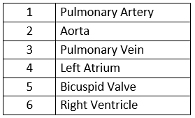
(d) Part 1 (Pulmonary artery) Deoxygenated blood Part 2 (Aorta) Oxygenated Blood
(e) In this phase, two valves, the bicuspid and tricuspid, are closed.
Question 8: Study the following diagram carefully:
(a) With which figure in the chapter can you roughly compare this diagram?
Write any two things shown in it as extra from those in the figure named above.

Solution 8:
(a) Diagrammatic relationship of artery, capillary and vein.
(b) The following are the two additional items displayed:
(i) Blood flow to and from the heart is depicted by arrows.
(ii) A capillary with red blood cells running through it.
Question 9: Given below is a diagrammatic representation of certain types of blood vessels in human body.
(a) Identify the types of blood vessels numbered 1 to 5.
(b) Where can such an arrangement be found as an example – in lungs or in heart walls?

Solution 9:
a)
1 – Arteriole
2 – Artery
3 – Venule
4 – Capillaries
5 – Vein
b) In the lungs, such an arrangement can be seen.
Question 10: Study the following diagram carefully and then answer the questions that follow:
(a) Name the cell labeled 1.
(b) Identify the phenomenon occurring in A.
(c) Mention two structural differences between 1 and 2.
Name the process occurring in B and C and state the importance of this process in the human body.

Solution 10:
(a) 1 – Red blood cell
(b) Diapedesis
(c)
| RBC | WBC |
| They don’t have a nucleus. | There is a nucleus in them. |
| They’re disc-shaped and biconcave. | They are round and come in a variety of sizes. |
(d) Phagocytosis is the process that occurs in B and C. The WBCs envelop and kill the foreign particles in this process, preventing disease from occurring.